<< Back to MOTIFvations Blog Home Page
DNA Methylation in Cell-free DNA (cfDNA): Benefits, Limitations & Future Potential for Precision Medicine
By Anne-Sophie Ay-Berthomieu, Ph.D.
November 2, 2021
Table of Contents:
Introduction
For a long time, disease diagnosis and prognosis have been performed using tissue biopsies, an invasive and often painful experience for a patient. Moreover, tissue samples only represent in-situ pathology, whereas, in cancer, metastatic cells can have a completely different biological profile as compared to the in-situ tumor. In this kind of disease, early detection and treatment are of crucial importance for increasing patient survival and decreasing relapse.
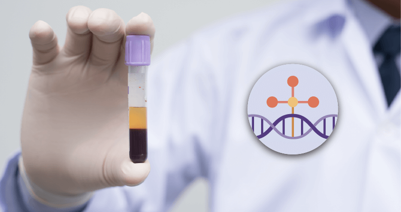
In recent years, liquid biopsies have sparked interest because collecting blood and urine samples is painless for the patient and technically easy to get. Besides the usual blood and urine analysis (metabolites, PBMC, etc.), scientists are interested in studying cell-free DNA (cfDNA), including quantity, sequence, and methylation status.
In this article, we look at what is cfDNA, its underlying benefit as a sample type, and the limitations of using such DNA for diagnosis and prognosis.
What is Cell-free DNA?
cfDNA – the History of Discovery
In 1948, Mendel and Métais detected DNA for the first time in human blood, but this discovery did not spark interest in the scientific community at the time. Fast forward to 1966, when Tan et al. demonstrated that cfDNA in the blood of patients suffering from systemic lupus erythematosus (SLE) leads to development of anti-dsDNA antibodies. In 1977, the first correlation between high cfDNA concentration and cancer was highlighted by Yaros’ lab. It was only in 1989 that Maurice Stroun and Philippe Anker demonstrated that in cancer patients, cfDNA originates, in part, from tumors. In 1994, a RAS point mutation was found in both cfDNA and tumor cells, confirming the origin of cfDNA.
Another interesting field for cfDNA detection and analysis is for monitoring pregnancy, beginning with the discovery in 1997 by Lo et al. that in pregnant women, cfDNA also comes from the fetus. cfDNA analysis allows the detection of Down syndrome, and determination of the sex and the Rhesus factor status of the fetus, without amniocentesis.
Shedding Happens
cfDNA is now known to have several origins. It is released by cells following apoptosis, necrosis, or via active secretion, providing a glimpse into the genomic and epigenomic state of the cells of origin. We have learned that cfDNA is predominantly comprised of double-stranded (ds) genomic DNA and mitochondrial (mt) DNA. Most cfDNA is found in oligonucleosome form and/or at the surface and the lumen of vesicles. Exosomes and chromatin folding itself can protect cfDNA from nuclease degradation in the blood.
The average size of cfDNA is 166bp, but can be up to 20-30kb, corresponding to mono and oligonucleosomes. This characteristic size is typical of caspase-dependent cleavage that occurs during apoptosis. Longer fragments typically come from necrotic cell death, while shorter cfDNA fragments are most often representative of circulating tumor DNA (ctDNA), mtDNA, and bacterial DNA.
The size distribution of cfDNA reflects cfDNA integrity. The ratio of long to short fragments and is significantly increased in cancer patients as compared to healthy patients and is also associated with tumor aggressiveness. Even without any further analysis, cfDNA fragment size determination itself is highly informative.
cfDNA can also be released by active secretion from live cells. For instance, erythroblasts secrete their nuclei during cell maturation, and neutrophils may also do so in response to various stimuli (NETosis). However, how, or why the cells get rid of their DNA and how it compensates for this loss is still not determined.
Liquid Biopsy - Advantages, Advantages, and More Advantages
The reason that cfDNA analysis is of great interest is because of the ease of blood sampling. The procedure is not invasive for the patient, is simple to perform, and the materials needed for collection are not expensive, allowing repetitive measurement during an experiment.
The second big advantage is the half-life of cfDNA, which ranges from 4 minutes up to 1-2 hours. cfDNA degradation is carried out by DNaseI, Factor H, and plasma Factor VII (FVII)-activating protease (FSAP) in the liver, kidneys, and spleen. The liver is doing most of the work: in 10 minutes 80% of free nucleosomes are cleared. In healthy patients, cfDNA clearance is fast, whereas, in patients with cancer or chronic inflammation, the increased level of cell death in their tissues overloads this clearance system, resulting in cfDNA accumulation. The short cfDNA half-life is convenient for real-time monitoring, including treatment response assessment which is a key factor in the field of precision medicine.
cfDNA Sample Prep – How Do We Begin...
Once liquid biopsy samples are collected, there is some challenge to cfDNA extraction due to the low yield of cfDNA in the plasma (1-100ng/mL of plasma), as well as the issue of possible contamination from genomic DNA (gDNA) from PBMCs.
Most of the time, blood is collected into an EDTA tube and then centrifuged to separate plasma and remove cell debris and gDNA contamination. Some collection tubes are specifically designed for cfDNA study, containing an additive that protects cfDNA from degradation and preventing gDNA release from PBMCs. Plasma preparation can impact the release of cellular nucleic acids and cfDNA modifications. Because of cfDNA’s short life, it should be extracted as soon as possible after plasma separation. Alternatively, plasma can be frozen at -80°C.
cfDNA Extraction
Many different kits are available to extract cfDNA from plasma. Most use columns or magnetic beads that bind to small size fragments corresponding to cfDNA size. Input volume is very important, and one should consider the quantity of cfDNA in the sample. In healthy patients, cfDNA concentration is very low whereas, in advanced cancer patients, it is higher. It is recommended to work with at least 1-2mL of plasma.
The first step in many of these protocols is pre-enrichment, where beads or columns bind small fragment DNA and are then washed, so that PBMC and gDNA are removed. The second step is elution of cfDNA from beads or columns so that cfDNA is collected in microliters of the elution buffer. cfDNA concentration is very low after extraction and devices such as Nanodrop are not sensitive enough to provide accurate quantitation of the sample. Instead, it is recommended to use a method such as the Qubit™ dsDNA High Sensitivity Assay for this purpose.
Active Motif has developed a cfDNA Purification Kit for preparation of cfDNA from 100µL to 10mL of serum and using a bead-based purification protocol that is compatible with automation.
Methods to Analyze DNA Methylation in Cell-free DNA
Another advantage of analyzing cfDNA is that it reflects the gDNA from healthy and damaged tissues as well as the epigenome, including DNA methylation (the addition of a methyl group (CH3) on position 5 on the pyrimidine ring of cytosine (5-mC)). DNA methylation is known to inhibit transcription when located in CpG islands, and to stabilize the genome when localized in non-coding regions. In cancer, we observe a global hypomethylation of the genome with hypermethylation of CpG islets, leading to genome instability and transcription repression, respectively. Thus, cfDNA methylation status could be a potential cancer biomarker. Different techniques are available to analyze the methylome, some of them are suitable for discovery research, whereas others can be adapted for clinical purposes.
Global Methylation ELISA
Global Methylation ELISA assays are useful in that they can reveal the percentage of DNA methylation or hydroxymethylation in a sample. However, the methylated sequences themselves are not specifically identified. This protocol consists of immobilizing DNA in a plate and measuring methylation or hydroxymethylation with a specific antibody. Quantification is performed with a standard curve, as in standard ELISA assays, which are well characterized, robust, low-cost, and sensitive.
Active Motif offers the Global DNA Methylation Assay–LINE-1 that measures the methylation level of LINE-1 repeats. It has been shown the LINE-1 methylation level reflects the whole genome methylation. This kit can detect 0.5% of methylation from 10 ng of DNA and perfectly works for cfDNA. Another version of the kit exists for 5-hydroxymethylcytosine quantification.
Bisulfite Conversion-based Methods
Bisulfite is a chemical that deaminates unmethylated cytosine to uracil, which is not found in gDNA. Methylation protects cytosines against deamination, so that 5-mCs are not converted to uracil. Following bisulfite conversion, DNA can be sequenced to single-nucleotide resolution. However, some limitations are that 5-mC and 5-hmC cannot be discriminated in this approach, and that DNA can undergo some degradation.
Whole-Genome Bisulfite Sequencing (WGBS) is the most comprehensive DNA methylation profiling. For cfDNA, sequencing adapters are bound to DNA fragments before conversion. WGBS is very expensive when producing high depth data.
A more cost-effective alternative is Reduced-Representation Bisulfite Sequencing. (RRBS) Using MspI digestion before conversion, sequencing is focused on CpG islands. (≈83%) RRBS does not cover intergenic regions and distal regulatory elements and covers regions only at proximity to the restriction site. For cfDNA samples, scRRBS was developed with low DNA input in a single-tube reaction.
Once the regions of interest are identified, Bisulfite Conversion can be followed by targeted PCR and sequencing of this region of interest. Primer design can be challenging. Nonetheless, this kind of strategy can be very interesting for diagnostic/prognostic use because it is less expensive, in particular the sequencing aspect, and is very sensitive (5-10ng of cfDNA).
Another solution is to use a methylation array, which contains predesigned probes covering 96% of CpG islands. Despite the low-genome-wide coverage, the low cost and the user-friendly protocol make microarrays a popular method.
Enrichment-based Approaches
The goal of enrichment-based methods is to concentrate methylated DNA to facilitate its detection, however, these techniques can have low resolution (100-300bp) and may display a bias towards hypermethylated regions.
MeDIP and hMeDIP use 5-mC and 5-hmC antibodies, respectively, to immunoprecipitate methylated or hydroxymethylated DNA fragments for sequencing. MeDIP-seq has been adapted for cfDNA (cfMeDIP-seq) requiring as little as 1-10ng of DNA. In this method, lambda DNA is used as a “filler” to increase the initial DNA input and maintain the mandatory DNA/antibody ratio. Lambda DNA does not have adapters, preventing it from being amplified and sequenced itself, so that the final DNA analyzed only comes from the original sample. As with most assays, the quality of the antibodies used is crucial to ensure accurate immunoprecipitation.
MBD-seq (methyl-CpG binding domain) is a technique that takes advantage of the biological property of certain proteins, including MBD2 and MeCP2, to recognize and bind to methyl-cytosine. It overperforms in regions with high CpG density as compared to MeDIP-seq. By adjusting the beads/DNA ratio and increasing incubation time, MBD-seq has been adapted to cfDNA, providing yet another approach to utilizing cfDNA for high throughput analysis.
hMeDIP can be used for 5-hmC identification, however, the sensitivity needed for this assay, combined with the lower yield of cfDNA from samples, is generally not optimal. In the more recently developed 5-hmC Seal, β-glucosyltransferase introduces an azide-modified glucose, that is then biotinylated. Biotinylated 5-hmC can then be concentrated using streptavidin beads and then sequenced with NGS. This technique is very sensitive and can work with as little as 5ng of cfDNA, but does have a limitation in that it cannot provide single-nucleotide resolution.
Restriction Enzyme-based Methods
Methylation-sensitive restriction enzymes (MRE) cleave only unmethylated sites, as methylation plays the role of protector against the activity of these enzymes. Taking advantage of this property, MRE-seq was developed. After digestion, DNA fragments are size-selected for library preparation. The protocol is quite easy to implement, however the coverage of the methylome is very weak due to limited CpG-containing cleavage sites. Additionally, the fragmented nature of cfDNA somewhat limits the size-selection post-digestion. MRE-seq was adapted to cfDNA with droplet digital PCR (ddPCR). In these methods, after digestion the methylation level is quantitatively determined on the microfluidic chip using real-time PCR. The ddPCR is ultra-sensitive and can work with 6.5-10 ng of DNA.
Discoveries enabled by cfDNA methylation studies
Renal Cell Carcinoma: cfDNA Hypermethylation & Tumor Stage Correlation
Renal Cell Carcinoma (RCC) is a deadly cancer, often found coincidentally with radiography. No reliable marker has been discovered as yet, and late diagnosis may lead to a rapid spread of this cancer. RCC patients do display an elevated level of cfDNA, which is a hallmark of the disease. Part of this cfDNA is circulating-tumor (ct) DNA, which may reflect the makeup of the in-situ tumor. S. Hauser et al. conducted a prospective study on sera from 35 RCC patients, including 15 with who had undergone radical nephrectomy and 20 with nephron-sparing surgery. The sera were collected just before surgery and the methylation profile of cfDNA from each was compared to sera from healthy patients.
To detect methylation, they used MRE-based assay to concentrate methylated cfDNA, followed with PCR. They targeted 8 genes involved in renal carcinogenesis: APC, GSTP1, p14(ARF), p16, RAR-B, RASSF1A, TIMP3, and PTGS2. In more than a half of patients, TIMP3 and APC genes were methylated, whereas methylation of p14 and GSTP1 was rarely found - 14.3% and 17.1%, respectively. Nevertheless, all the genes were hypermethylated in RCC patients as compared to healthy patients, except for p16 and TIMP3. Increased APC methylation was found to correlate with locally advanced disease in patients, however no additional correlation was detected with other clinico-pathological parameters.
This kind of cfDNA analysis could be an interesting diagnostic/prognostic tool for RCC detection and treatment.
5-Hydroxymethyl-cytosine as a Potential Biomarker for Esophageal Cancer
Esophageal cancer is the 8th most common cancer worldwide and the 6th leading cause of cancer death. Like for many cancers, an early diagnostic is the key to recovery. X. Tian et al. analyzed the hydroxymethylome of cfDNA from 150 esophageal cancer patients. They used the nano-hmC-Seal method to concentrate 5-hmC DNA fragments and sequenced them to identify the genes involved. They observed that more than 9000 5-hmC-gain regions and more than 10000 5-hmC-loss regions. In cancer patients, 5-hmC-gain regions were mostly located in gene bodies, including promoter and UTR regions. They were also enriched in short interspersed nuclear elements, long tandem repeats, and satellite repeats. Altogether, these observations suggest that the 5-hmC profile is significantly different between healthy and cancer patient cfDNA.
For genes differentially hydroxymethylated in cancer, they also showed that esophageal cancer samples displayed a distinct signature from healthy samples. Functional enrichment analysis showed that carcinogenesis-related pathways such as Hippo, PI3K-Akt, and MAPK signaling were enriched. By comparing the 5-hmC profiles in esophageal cancer to previous studies in colorectal and gastric cancer, they observed that the signature is unique to esophageal cancer.
Finally, they used the 5-hmC profile for cancer classification and were able to generate a model, using 5 markers, to classify esophageal cancer stages. This study provides a solid basis for the development of diagnostic tools from body fluids, including analysis of cfDNA.
Colorectal Cancer Screening Using the cfDNA Epigenome
Colorectal cancer (CRC) is the 4th most cancer-related death. Diagnosis necessitates patients undergo an invasive coloscopy procedure. L. De Chiara’s lab investigated the possibility of using cfDNA for CRC cancer diagnostic instead. They had access to 60 serum samples, 20 from healthy patients (NCF), 20 from patients with advanced adenomas (AA), and 20 from patients with colorectal cancer (CRC). They used a pooling sample method to evaluate cfDNA methylome with MethylationEPIC BeadChip (866836 CpGs), where two pools from 10 patients per pool were prepared.
Their analysis found that AA and CRC pools were globally hypomethylated as compared to the NCF group. Unsupervised clustering allowed differentiation between advanced neoplasia and healthy controls. They also observed that the methylation profiles of AA and CRC clustered close together.
Again, we see the value and first steps toward using cfDNA as a non-invasive procedure to detect CRC, without coloscopy.
cfDNA Methylation Profiling for Lung Cancer Diagnostic
Lung cancer is the 2nd most common cancer in men and women and is the 1st leading cause of cancer death. Genetic testing for early diagnosis is not currently available and the use of body fluid to detect such a deadly disease is very appealing as a screening tool. W. Xu et al. analyzed by MeDIP-seq the methylome of cfDNA from 5 patients with lung cancer and 3 healthy individuals. They found that the methylation pattern in lung cancer patients was significantly different from healthy patients. The majority of DNA methylated regions (DMR) of interest were in intergenic regions.
It is known that promoter hypermethylation is a hallmark of cancer. Interestingly, this study found that over 300 DMRs were located in promoter regions, 33 hypermethylated and 297 hypomethylated. Some genes with hypermethylated promoters are involved in lung tumorigenesis, such as GAS7, AQP10, HLF, and HOPX. Finally, to confirm that the 33 genes with hypermethylated promoters were involved in lung cancer, they analyzed publicly available DNA methylation data in 36 cancer patients and 36 healthy individuals. They identified 10 genes which were significantly hypermethylated, including B3GAT2, BCAR1, HLF, HOPX, HOXD11, MIR1203, MYL9, SLC9A3R2, SYT5, and VTRNA1-3. Altogether, these results suggest that cfDNA could be used as a diagnostic tool for lung cancer, using the methylation status of the 10 above-mentioned genes as a signature for screening.
Summary & Future
We know that cfDNA can be found in body fluids, including blood, urine, and feces, originating from the elimination of dead cells or secretory vesicles, and are believed to reflect the gDNA makeup of the tissue of origin.
Extraction can be challenging but many methods and commercially available kits are available, which capture cfDNA while avoiding cell DNA contamination, cfDNA degradation, and importantly, allowing automation. Moreover, sample collection is non-invasive and inexpensive.
cfDNA analysis offers a window into the body’s general state - where an increasing quantity of cfDNA suggests an increase in cell death associated with several pathologies such as cancer and auto-immune diseases. The sequence of cfDNA can also help to detect mutations linked to a specific disease, such as KRAS mutation in cancer. Finally, the methylation profile of cfDNA can be a useful tool for diagnostic and prognostic since DNA methylation modification is one of the first epigenetic modifications that occurs in the cells path to carcinogenesis.
To adapt cfDNA analysis for clinical purposes, much more research will be needed in order to hopefully identify unique signatures that can be correlated with specific disease(s). Whereas BWGS gives in-depth information about the cfDNA methylation status, strategies such as Targeted Bisulfite Sequencing may be more suitable for diagnostic applications as they are more cost-effective in the clinical setting.
Overall, cfDNA quantity and quality is a promising tool for diagnosis and prognosis purposes, and research in this regard has exploded in recent years, hoping to systematically identify and link cfDNA quantity, sequence, and/or methylation status to a particular disease.
About the author

Anne-Sophie Ay-Berthomieu, Ph.D.
Anne-Sophie was born in the south of France and grew up between the Mediterranean Sea and the Pyrenean Mountains. She grew up as a science fiction fan, leading her to specialize in molecular biology and genetics during graduate school at the University of Lyon, France (secretly hoping her research would give her superpowers!). After living in different places for work, she is back in Lyon, France where she shares her time between her husband, her family, and her friends. During her free time, Anne-Sophie challenges herself with hiking, climbing, racing, and traveling in foreign countries – while waiting for her superpowers to grow!
Contact Anne-Sophie on LinkedIn with any questions, or to tell her about your superpowers.
Related Articles
Epigenetics of Liquid Biopsies, Histone Ubiquitination, and piRNAs & SUMO
January 22, 2020
This latest epigenetics news summary covers approaches to investigate epigenetics and proteomics of liquid biopsies, a new role for histone ubiquitination in regulation of gene expression, and new insights into piRNA-mediated transcriptional silencing.
Read More
<< Back to MOTIFvations Blog Home Page


