For GSDIM (Ground State Depletion with Individual Molecule return) microscopy using the Leica SR GSD microscope, the use of fluorescent Rhodamine 6G dyes are strongly recommended by Leica Microsystems. The fluorescent properties of these dyes meet the specifications required to perform GSDIM microscopy with the 500 mW 532 nm laser-line nm laser of the SR GSD system. Active Motif's Fluorescent Secondary Antibody Conjugates have been prepared using an optimized protocol that ensures the highest fluorescent intensity and stability. In addition, the Rhodamine 6G conjugates have been maximally cross-adsorbed against IgG's of a variety of species to eliminate background caused by non-specific binding. These unique features make the Active Motif Rhodamine 6G secondary conjugates ideal tools for GSDIM microscopy.
To ensure that you achieve the best image quality possible, Rhodamine 6G (GSD)-stained slides should be embedded in either Glucose-Oxidase Mix (containing glucose, glucose oxidase and catalase in a PBS buffer of pH = 7.4) or Mowiol -based embedding media.
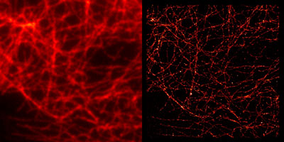
Figure 1: Active Motif Rhodamine 6G (GSD) antibody conjugates in widefield and GSDIM microscopy.Tubulin was stained with a primary monoclonal mouse antibody and with Rhodamine 6G (GSD) Goat anti-mouse IgG (Catalog No. 15074) secondary antibody. The widefield (left) and GSDIM (right) images are courtesy of Leica Microsystems, Germany.
For additional information on Rhodamine 6G (GSD) antibody conjugates, please visit the Fluorescent Secondary Antibody Conjugates page.