Cart
Cart Products
ChIC/CUT&RUN Assay Kit Overview
Related Products:
- CUT&RUN-validated antibody: For best results with your samples
- CUT&RUN Spike-In Control: Evaluate CUT&RUN assay datasets confidently for comparison
- DNA Library Prep Kit for Illumina®: Dual Index NGS Kit for ChIP-Seq, CUT&RUN, and ds methylated DNA assays
- Tissue Prep for NGS Assays: Tissue sample preparation for CUT&RUN
- Magnetic Separation Rack: Rapid and efficient magnetic bead-based separation of samples
- Diversi-Phi Indexed PhiX: Reliable diversity for every Illumina sequencing run
- CUT&RUN Workshops: Hands-on, in‑person workshops to advance your epigenomics expertise
CUT&RUN (Cleavage Under Targets & Release Using Nuclease) is an epigenetic method used to investigate the genome-wide distribution of various chromatin-associated proteins and their modifications1-3. CUT&RUN is a derivative of chromatin immunocleavage (ChIC). CUT&RUN is similar to chromatin immunoprecipitation (ChIP), in that it utilizes an antibody to target chromatin-associated marks and proteins, but requires less sample material and less sequencing depths than ChIP.
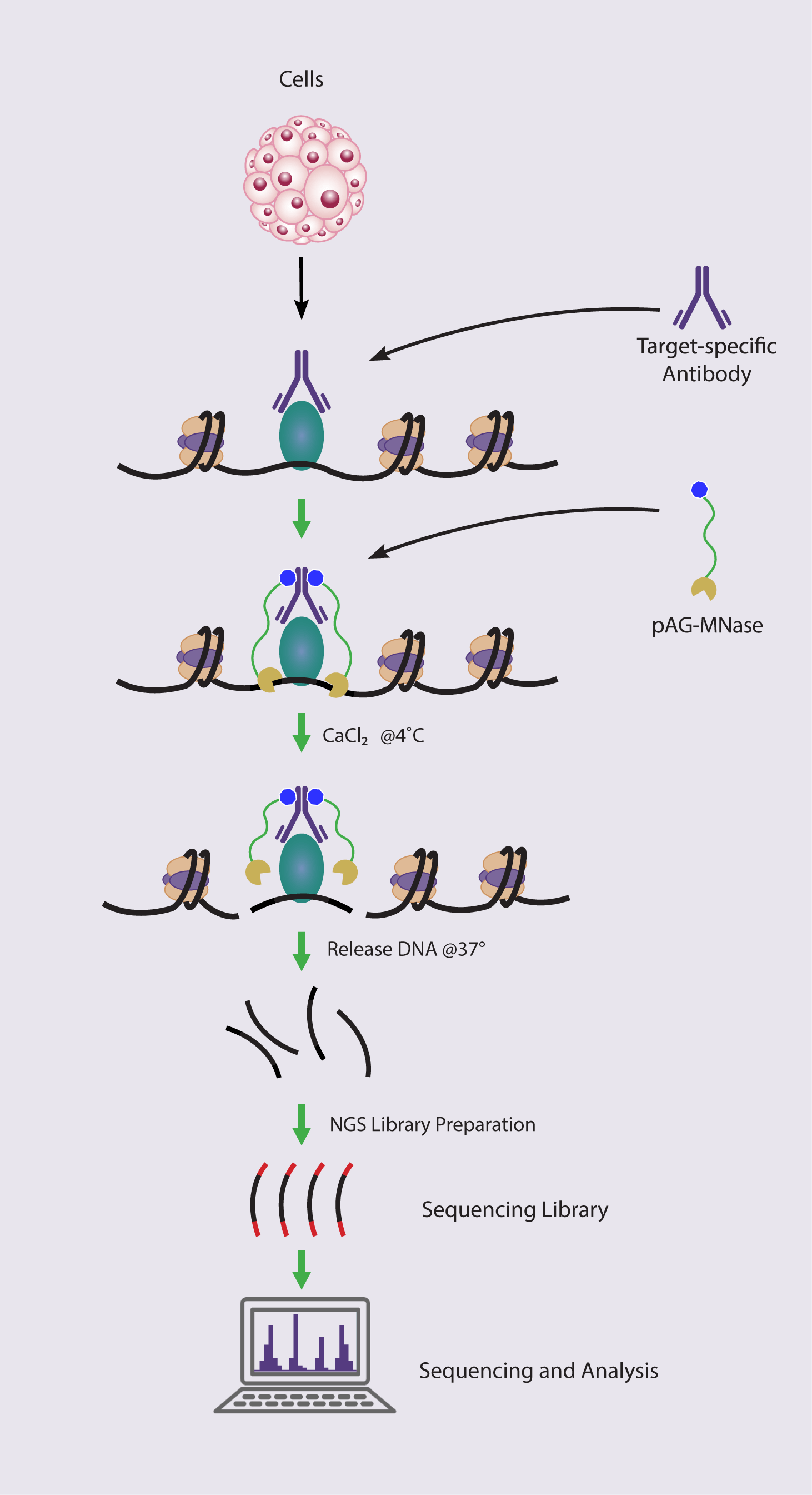
In CUT&RUN, a protein of interest is tagged with an antibody and bound to the chromatin in intact cells. Then, a micrococcal nuclease (MNase) is used to cleave the DNA specifically at the binding sites of the protein of interest. The released fragments are purified, sequenced, and mapped to the reference genome to determine the protein’s binding sites. Unlike ChIP, CUT&RUN does not require crosslinking of the protein to the DNA, which can introduce artifacts.
CUT&RUN is a valuable tool for studying chromatin-associated proteins because it is sensitive, specific, and requires fewer cells than ChIP, making it ideal for identifying binding patterns of chromatin-associated proteins such as transcription factors or histone modifications genome-wide. Chromatin-associated proteins play critical roles in regulating various cellular processes such as gene expression, DNA replication, DNA repair, and cell differentiation. Understanding the binding patterns of these proteins can provide insight into how these cellular processes are regulated.
References:
1. Schmid, M. et al. Mol Cell, 16(1): 147-157 (2004)
2. Skene, P.J. et al. (2017) Elife 6, e21856
3. Skene, P.J. et al. (2018) Nat. Protoc., 13, 1006-1019
ChIC/CUT&RUN Assay Kit Advantages:
- Compatible with 5,000 to 500,000 cells
- Complete kit with optimized protocol
- Developed for genome-wide chromatin-associated protein profiling
How the ChIC/CUT&RUN Assay Works
CUT&RUN vs. CUT&Tag vs. ChIP-Seq
| CUT&RUN | CUT&Tag | ChIP-Seq | |
|---|---|---|---|
| Performed Under Native Conditions? | Yes | Yes | No |
| Chromatin Fragmentation Method | MNase digestion | Tn5-based tagmentation | Sonication |
| Cell Number Requirements | 500,000 cells | 5,000-500,000 cells | 1-10 million cells |
| Sequencing Depth Required * | 8 million reads † | 2 million reads ** | 20-50 million reads |
| Integrated Library Preparation? | No, separate library prep required | Yes, uses tagmentation | No, separate library prep required |
| Compatible Targets | Wide range of histone modifications, transcription factors, and co-factors | Primarily histone modifications, some transcription factors and co-factors | Wide range of histone modifications, transcription factors, and co-factors |
| Workflow Length | 1-2 days | 1-2 days | 2-3 days |
* Kaya-Okur et al. Nature Communications (2019) 10:1930
** For less abundant targets of interest, 8 to 10 million reads are recommended
† 25 million reads are recommended for transcription factor targets

ChIC/CUT&RUN Assay Kit Contents
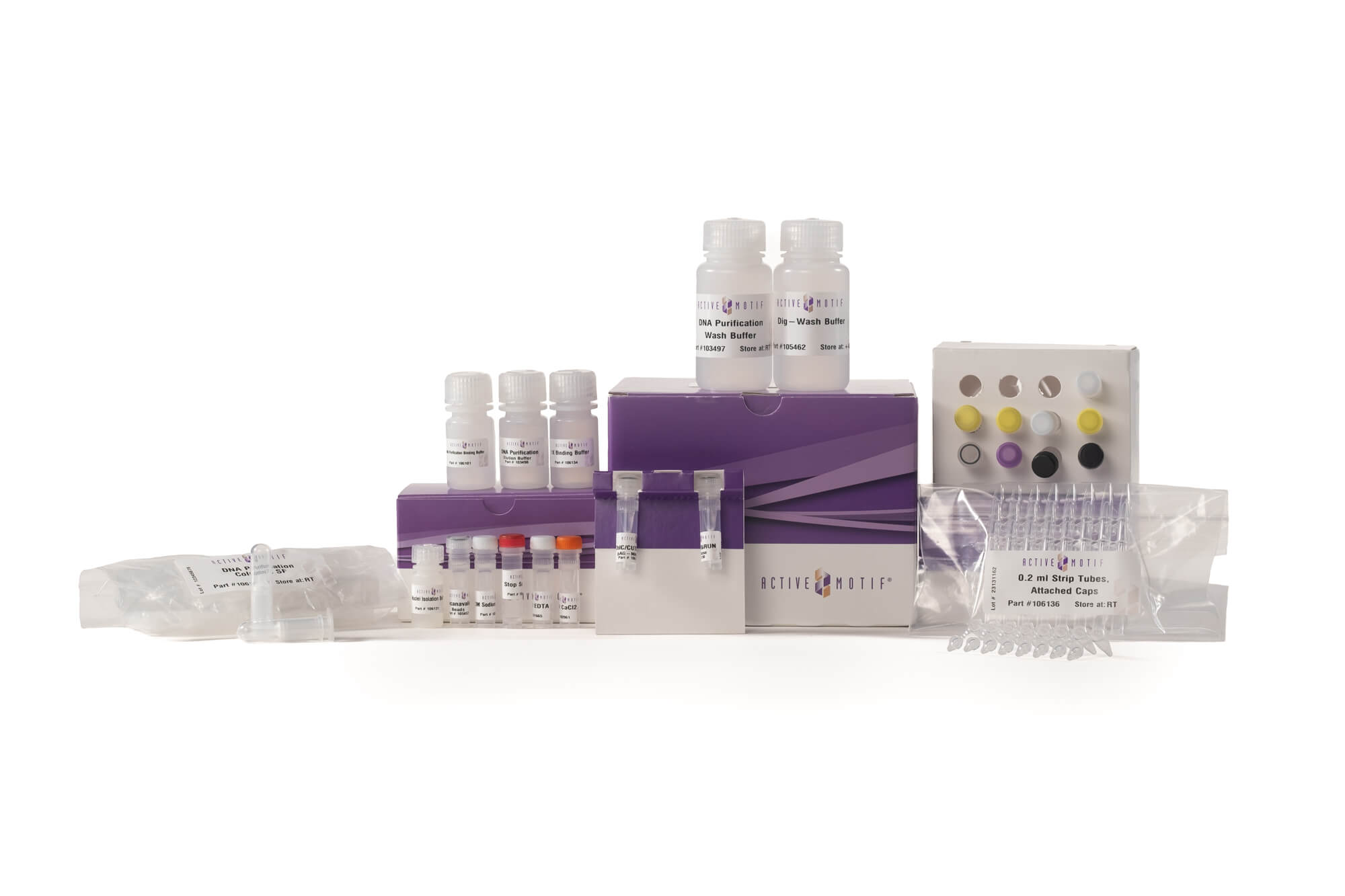
The ChIC/CUT&RUN Assay Kit components are shipped at two temperatures, with one box on dry ice for components to be stored at -20°C, and a second box at room temperature for components that are not to be frozen and must be stored at 4°C. Please store components according to the storage conditions in the manual after first use. All reagents are guaranteed stable for 6 months from date of receipt when stored properly.
- Protease Inhibitor Cocktail, 500 µl, store at -20°C
- ChIC/CUT&RUN pAG-MNase, store at -20°C
- Glycogen 20 mg/ml, store at -20°C
- Histone H3K4me3 pAb, 10 µg, 1 mg/mL store at -20°C
- Negative Control IgG, Rabbit, store at -20°C
- 1 M Spermidine, store at -20°C
- Nuclei Isolation Buffer, store at 4°C
- Concanavalin A Beads, store at 4°C
- 0.1 M CaCl2, store at 4°C
- Stop Solution, store at 4°C
- 1X Binding Buffer, store at 4°C
- Dig-Wash Buffer, store at 4°C
- 0.5 M EDTA, store at Room temperature
- DNA Purification Wash Buffer, store at Room temperature
- DNA Purification Elution Buffer, store at Room temperature
- DNA Purification Binding Buffer, store at Room temperature
- 3M Sodium Acetate, store at Room temperature
- DNA Purification Columns SF, store at Room temperature
- 0.2 mL Strip Tubes, Attached Caps, store at Room temperature
- RNase A, store at -20°C
- 5% Digitonin, store at -20°C
ChIC/CUT&RUN Assay Kit Data
(Click image to enlarge)
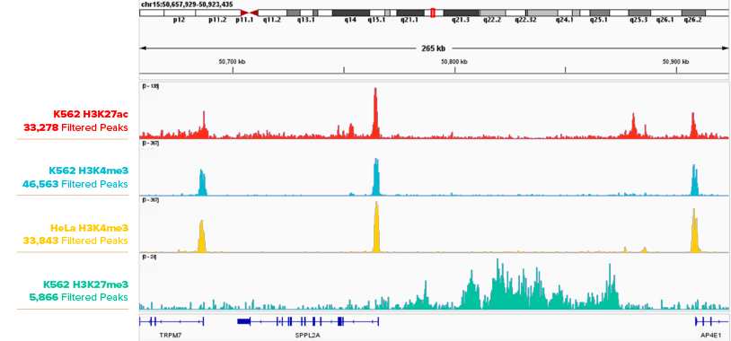
Figure 1. CUT&RUN Profiling Histone Marks in K562 Cells.
IGB browser tracks are shown for 500,000 K562 cell CUT&RUN peaks with Active Motif antibodies H3K27ac (Cat. No. 91193), H3K4me3 (Cat. No. 91263) and H3K27me3 (Cat. No. 39155).
 (Click image to enlarge)
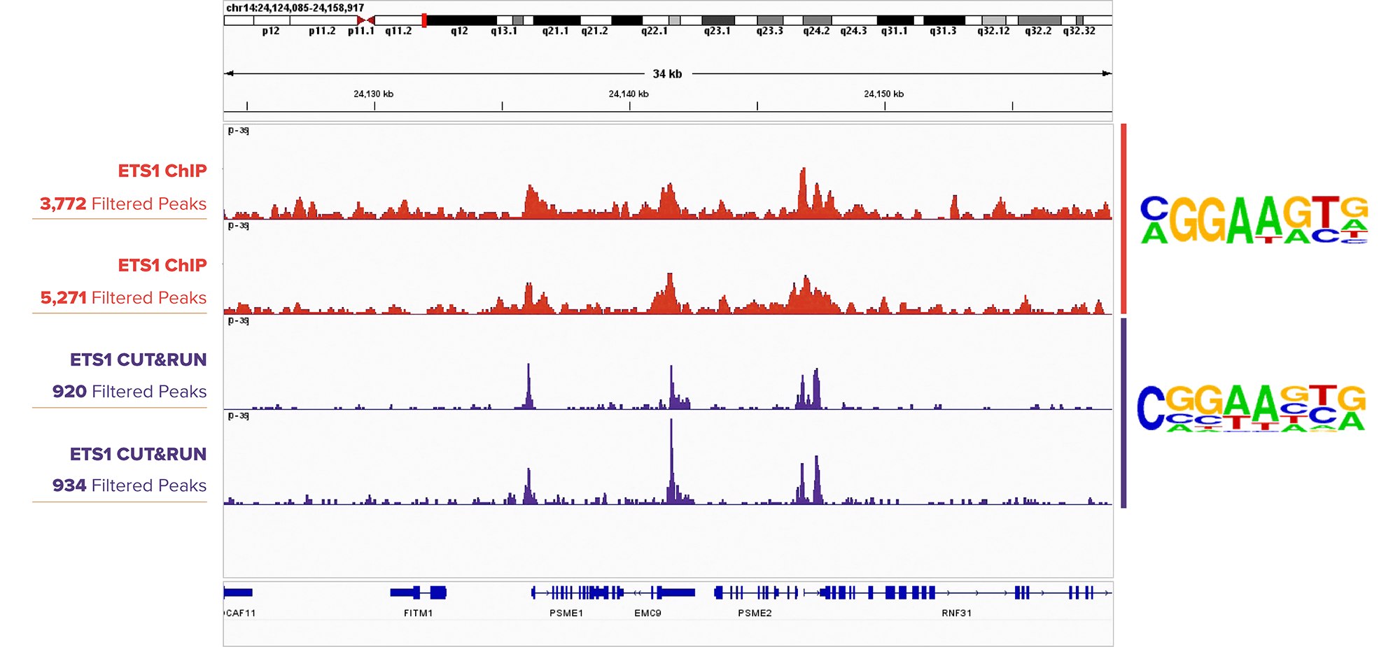
Figure 2. ETS1 CUT&RUN Peak Profiles Are Similar to ETS1 ChIP-Seq Profiles.
IGB browser tracks for ChIP-Seq (top two tracks) in DIPTG cells and CUT&RUN (bottom two tracks) in K562 cells assays targeting ETS1 (Cat. No. 39580). Sequence logos from Homer analysis (to the right of the tracks) show motif enrichment for the ETS1 motif in both sample sets.
 (Click image to enlarge)
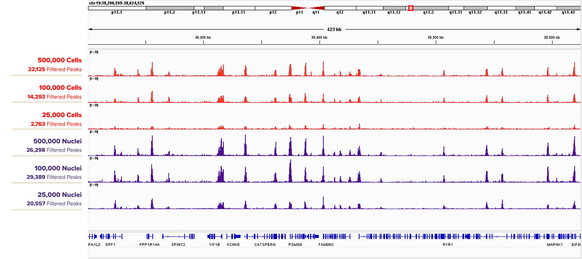
Figure 3. CUT&RUN H3K4me3 Assay Results Comparing Titrations of K562 Cells versus Nuclei.
500,000, 100,000, 25,000 K562 Cells (top three tracks) or Nuclei (bottom three tracks) were assayed in CUT&RUN with Active Motif’s H3K4me3 antibody (Cat. No. 39060). Filtered Peaks remain higher at lower input for nuclei than cells, and the browser tracks for nuclei show stronger peaks for each input amount when compared to cells.

(Click image to enlarge)
Figure 4. Active Motif’s CUT&RUN Assay Kit Outperforms the Competition.
Peak scores are plotted in the bar graphs for 500,000, 100,000, 25,000, and 5,000 K562 nuclei assayed in CUT&RUN Kits from Active Motif and Competitor for YY1 (Active Motif Cat. No. 61980) and H3K4me3 (Active Motif Cat. No. 39916).

(Click image to enlarge)
Figure 5. Active Motif’s CUT&RUN Assay Kit is Compatible with 5,000 Nuclei for Histone Marks.
5,000 fresh K562 nuclei were assayed using Active Motif’s CUT&RUN Assay Kit and Competitor CUT&RUN Assay Kits using Active Motif's H3K4me3 antibody (Cat. No. 91263). Active Motif’s CUT&RUN Assay Kit yielded more robust results than the competition.

(Click image to enlarge)
Figure 6. Active Motif’s CUT&RUN Assay Kit is Compatible with 25,000 Nuclei for Transcription Factors.
25,000 fresh K562 nuclei were assayed using Active Motif’s CUT&RUN Assay Kit and Competitor CUT&RUN Assay Kits using Active Motif's YY1 antibody (Cat. No. 61980). Active Motif’s CUT&RUN Assay Kit yielded more robust results than the competition.
ChIC/CUT&RUN Assay Kit FAQs
1. Sample Type, Input & Experimental Design
For transcription factor targets, we recommend using approximately 500,000 cells per CUT&RUN reaction. Using a CUT&RUN-validated antibody is strongly recommended to ensure specific binding and robust signal.
- Recommended input: ~500,000 cells per reaction
- Target type: Transcription factors
- Antibody requirement: CUT&RUN-validated antibody
Related: CUT&RUN-Validated Antibodies
For most applications, isolated nuclei are recommended for CUT&RUN because they generally produce cleaner signal and higher specificity. However, intact cells can yield comparable or, in some cases, higher peak profiles depending on the target.
- General recommendation: Use nuclei for consistent, low-background results
- Transcription factors: Some targets show similar or higher peaks in intact cells
- Histone marks: Nuclei typically produce stronger and more consistent enrichment
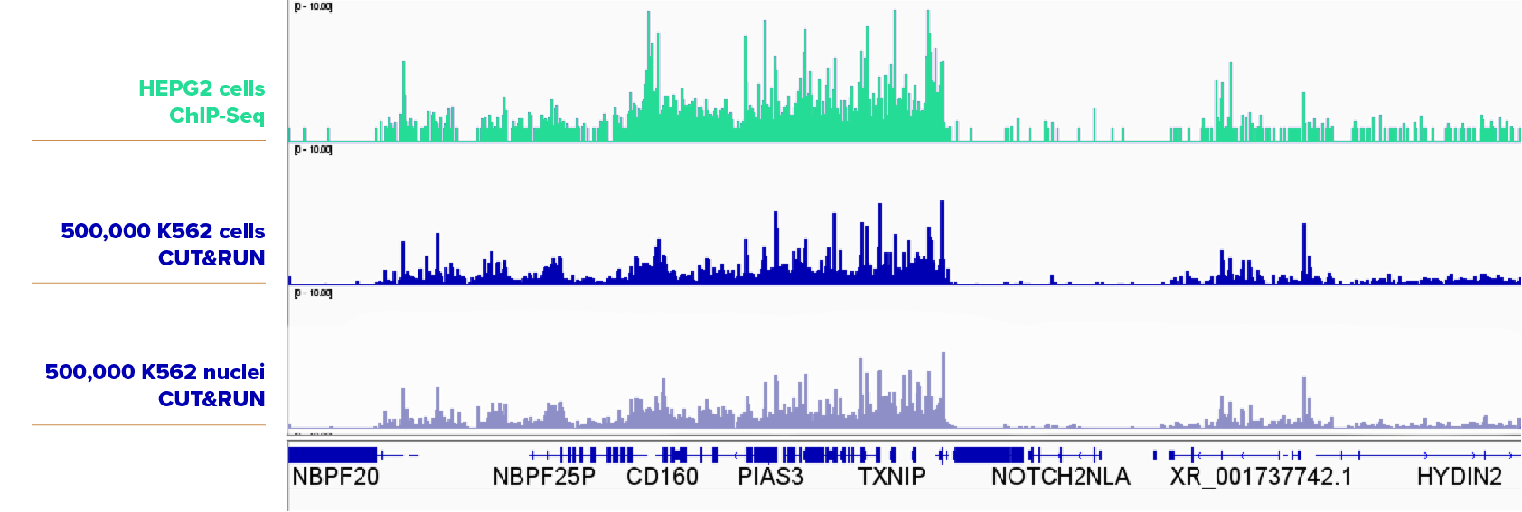
The examples below compare CUT&RUN results generated from equal numbers of K562 cells and K562 nuclei using validated antibodies.
(Click image to enlarge)
Using 500,000 K562 cells or nuclei with the SUZ12 antibody (Cat. No. 39357), both sample types produced highly similar CUT&RUN profiles. ChIP-seq data from HEPG2 cells is shown for reference.

(Click image to enlarge)
For BRD family transcription factors, intact cells yielded slightly higher peaks for BRD2 (Cat. No. 61797) and BRD3 (Cat. No. 61489), while nuclei produced higher enrichment for BRD4 (Cat. No. 91301).
(Click image to enlarge)
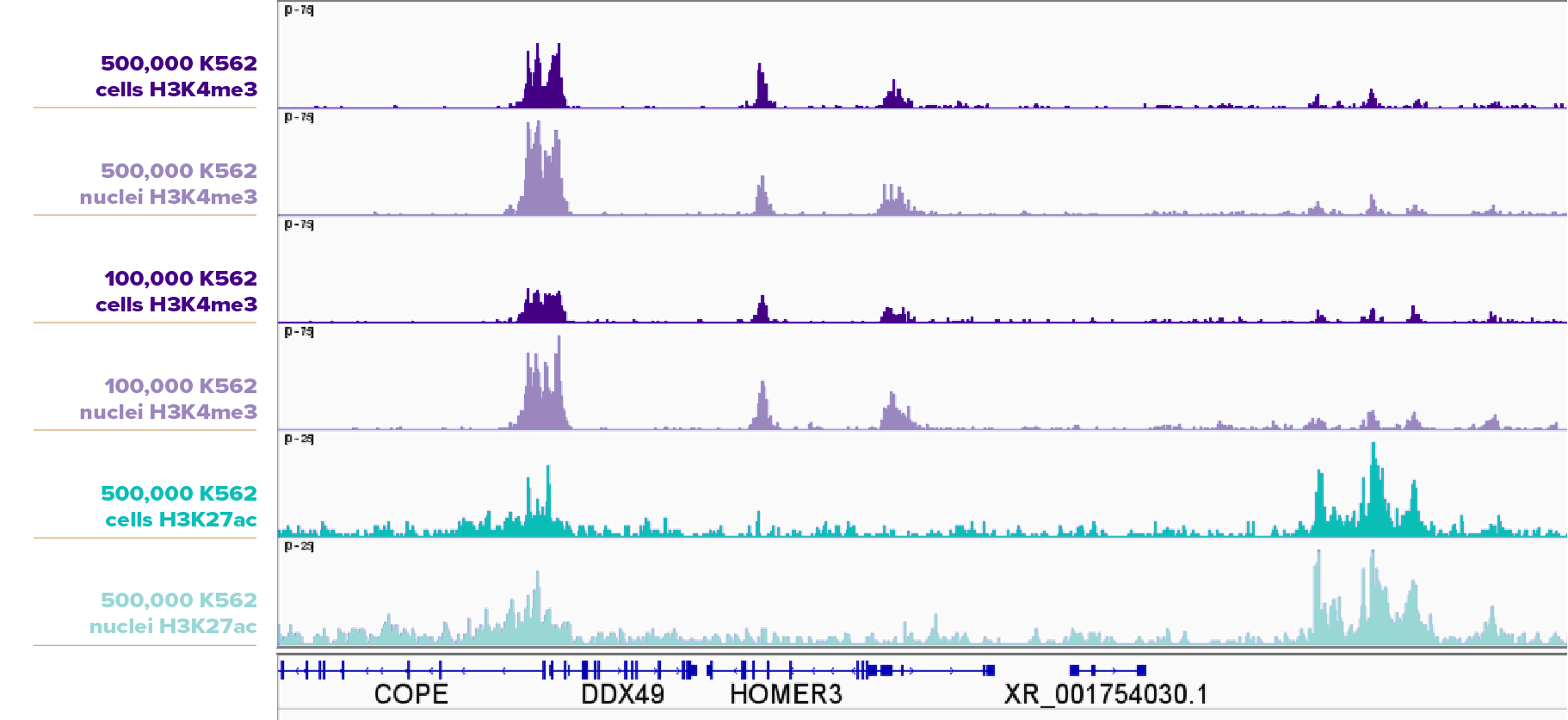
For histone modifications including H3K4me3 (Cat. No. 91263) and H3K27ac (Cat. No. 91193), nuclei consistently produced higher signal and sharper peaks than intact cells.
No, it is not required. Use a spike-in control when you need a more accurate comparison of signal intensity across samples. This is especially important for experiments involving treatments, genetic perturbations, or time courses where global chromatin changes may occur. Spike-ins also help account for technical variability such as differences in cell number, digestion efficiency, or sample quality. If your goal is purely to identify binding sites or peak locations within a single sample, a spike-in may not be necessary; however, for quantitative comparisons between conditions, it is strongly recommended.
Related: CUT&RUN Spike-In Control
Yes, any library prep kit validated for a small number of short fragments is acceptable. We validated 2 library prep kits for use this kit:
- DNA Library Prep Kit for Illumina® (Active Motif Cat. No. 53220)
- Dual Index Primers Set 1 for Illumina® (Active Motif Cat. No. 53221)
- Dual Index Primers Set 2 for Illumina® (Active Motif Cat. No. 53222)
- NEBNext® Ultra™ II DNA Library Kit for Illumina: E7645L
- Indexing primers such as NEBNext Multiplex Oligos for Illumina (Index Primers Set 1): E7335S
- Indexing primers such as NEBNext Multiplex Oligos for Illumina (Index Primers Set 2): E7500S
No, this kit has not been validated in plant samples. We suspect that plant samples can be used with the kit if high quality nuclei can be isolated using a validated nuclei isolation protocol for your specific sample type.
2. Beads, Pellets & Physical Workflow Checks
Yes, the nuclei pellet may still be present even if it is not visible. When using the CUT&RUN Kit, nuclei pellets can be very small or translucent. Always orient tubes consistently in the centrifuge so you know where the pellet should form, and carefully remove supernatant without disturbing the pellet.
- Pellet visibility: May be faint or invisible
- Best practice: Keep tube caps oriented in the centrifuge
- Handling tip: Proceed slowly when aspirating supernatant
Related: CUT&RUN Kit Manual
Properly prepared Concanavalin A (Con A) beads appear uniform and opaque, and cell-bound beads form visible aggregates or clusters. During the CUT&RUN workflow, successful cell binding is indicated by beads that pellet efficiently and show increased opacity due to attached cells.
- Unbound beads: Uniform, opaque suspension
- Cell-bound beads: Visible clumping or heavier pellet formation
- Best practice: Gently mix to avoid bead loss or uneven binding
Watch: This short video demonstrates how to prepare Con A beads for CUT&RUN or CUT&Tag and what to look for during bead binding.
If SPRI beads are slow to pellet, gradually guide them using a handheld magnet before placing the tubes on a magnetic plate. Begin by pelleting the beads toward the top of the tube, then slowly move the magnet downward to collect the beads at the bottom.
- First step: Use a handheld magnet to move beads from top to bottom
- Final step: Place tubes on a magnetic plate to fully pellet beads
- Tip: Avoid disturbing beads once pelleted to prevent loss
3. Antibodies & Target Compatibility
The antibodies listed below have been used in published CUT&RUN studies. These antibodies were cited in peer-reviewed publications but have not been formally validated for CUT&RUN by Active Motif.
- Use case: Reported in published CUT&RUN experiments
- Validation status: Not officially CUT&RUN-validated by Active Motif
- Recommendation: Independent optimization may be required
Related: CUT&RUN-Validated Antibodies
Reference list: Browse antibodies reported in CUT&RUN publications below.
Cookies are required to display this table. Click here to reopen the cookie settings and enable non-essential cookies.
Not necessarily. Antibodies validated for ChIP-seq or CUT&Tag do not always perform well in CUT&RUN because each method uses a distinct workflow and binding context.
- ChIP-seq: Relies on crosslinking and sonication
- CUT&Tag: Uses tethered tagmentation
- CUT&RUN: Requires efficient antibody binding under native conditions for pA-MNase cleavage
For best results and reproducible data, we recommend using Active Motif CUT&RUN-validated antibodies, which have been tested specifically for performance in the CUT&RUN Assay Kit.
Recommended: CUT&RUN-Validated Antibodies
4. Controls, Background & Troubleshooting
High background in CUT&RUN data is most commonly caused by poor cell or nuclei quality. For optimal results with the CUT&RUN Kit, start with highly viable cells and intact nuclei to minimize nonspecific cleavage.
- Check viability: Use automated counters (e.g., Countess II) or Trypan Blue exclusion
- Assess integrity: Confirm nuclei are intact before proceeding
- Best practice: Avoid harsh handling that can damage cells or nuclei
Yes, the negative control IgG will usually produce a sequencing library when using the CUT&RUN Kit. However sometimes there is not enough library to sequence. This low-level library reflects background cleavage and is expected, serving as an important reference for data normalization and quality assessment.
- Control type: IgG negative control
- Expected result: Library generation with low signal
- Purpose: Background measurement and data normalization
5. Library QC & Fragment Profiles
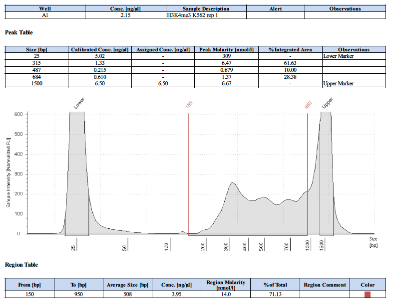
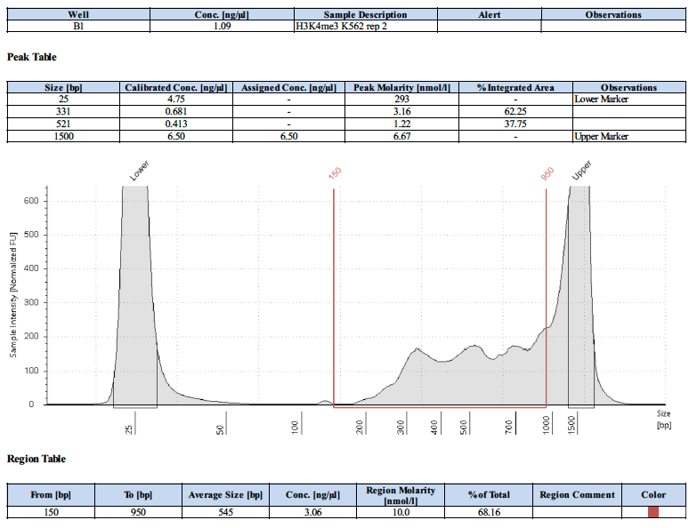
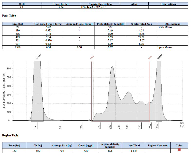
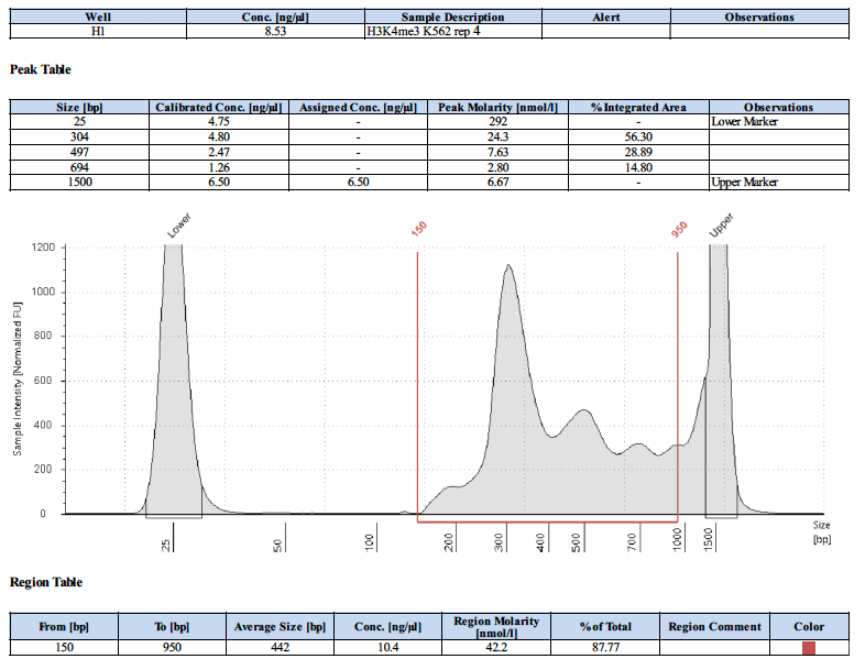
Positive control CUT&RUN libraries show a characteristic enrichment of short DNA fragments, typically peaking around mono- and sub-nucleosomal sizes (200-600bp). These fragment distributions indicate successful target-specific cleavage and library preparation.
- Expected fragment sizes: ~150 bp and smaller (after adapter removal)
- Signal quality: Distinct peaks with low high-molecular-weight background
- Interpretation: Confirms effective antibody binding and pA-MNase activity
- Learn More: Watch our video about library QC assessment here
Below are representative library trace examples generated using the CUT&RUN positive control antibody.
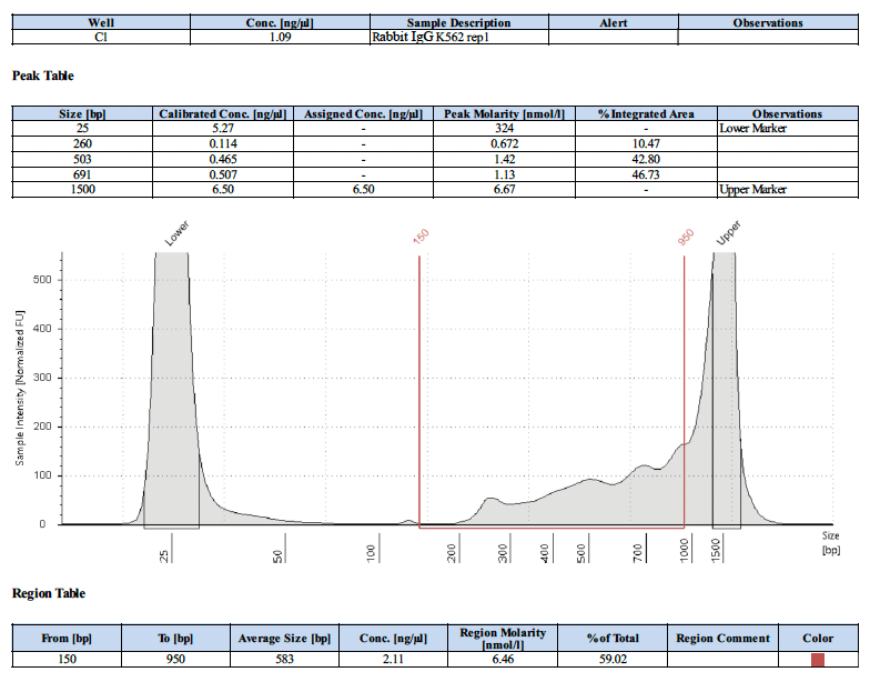
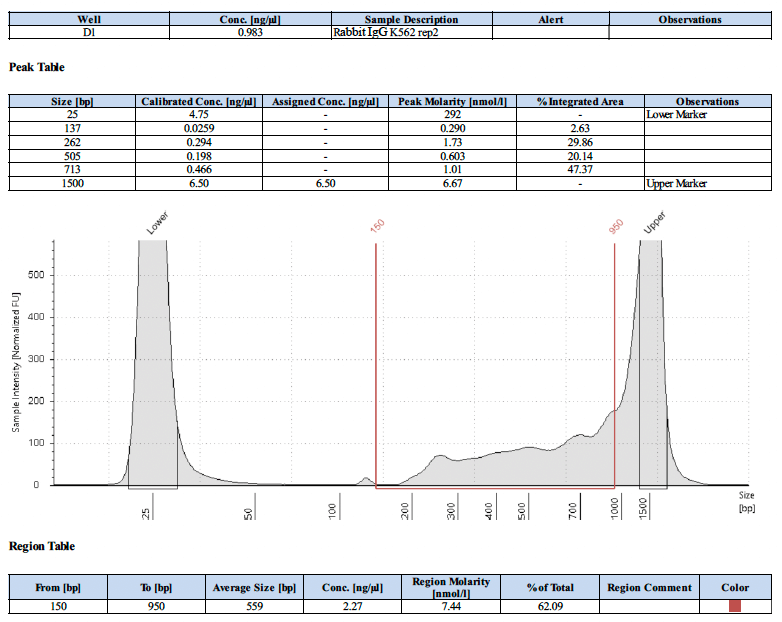
CUT&RUN negative control IgG libraries typically show low signal with a broad DNA fragment distribution and no distinct enrichment peaks. This pattern reflects background cleavage rather than target-specific binding. Often the IgG control does not produce enough library to sequence, so it is common to omit it from the analysis. For narrow marks, we typically avoid calling peaks directly against the IgG control, as this can sometimes be overly stringent. Generally, we call peaks first and then use the IgG as a visual reference to rule out spurious peaks or remove them via overlap.
- Expected fragment profile: Broad distribution without clear mono- or sub-nucleosomal peaks
- Signal intensity: Lower overall signal compared to positive control antibodies
- Interpretation: Confirms low nonspecific cleavage and assay specificity
- Learn More: Watch our video about library QC assessment here
Below are representative library trace examples generated using the CUT&RUN negative control IgG.
YY1 CUT&RUN libraries typically show a strong enrichment of short DNA fragments, consistent with transcription factor binding. The profile is dominated by sub-nucleosomal fragments, indicating efficient antibody targeting and precise pA-MNase cleavage.
- Expected fragment sizes: Primarily <150 bp (after adapter removal)
- Peak shape: Sharp, well-defined enrichment peaks
- Interpretation: Confirms successful CUT&RUN profiling of YY1 binding sites. YY1 is a widely expressed target and well characterized, so it makes a good positive control to run alongside a less abundant TF.
- Learn More: Watch our video about library QC assessment here
A representative library trace generated using a CUT&RUN-validated YY1 antibody is shown below.

JunD CUT&RUN libraries typically show strong enrichment of short, sub-nucleosomal DNA fragments, consistent with transcription factor binding. The fragment profile reflects precise pA-MNase cleavage at JunD binding sites.
- Expected fragment sizes: Predominantly <150 bp (after adapter removal)
- Peak shape: Sharp, well-resolved enrichment peaks
- Interpretation: Indicates successful CUT&RUN profiling of JunD occupancy. JunD is a good positive control target because it is highly expressed in numerous cancer cell lines and is well charactierized.
- Learn More: Watch our video about library QC assessment here
A representative library trace generated using a CUT&RUN-validated JunD antibody is shown below.

Yes, it is possible. However, we do not have a recommended protocol as we prefer NGS to validate the success of the CUT&RUN assay.
6. Sequencing Recommendations
Recommended sequencing depth depends on the target. Typically, 10 million reads for most targets. Usable results can sometimes be obtained with as few as 3-5 million reads for abundant histone markers. For low abundancy transcription factors, 20-30 million reads may be needed.
- Transcription factors: ~10-30 million reads per sample depending on abundance
- Histone marks: ~3-10 million reads (target-dependent)
- Note: Deeper sequencing may be required for low-abundance targets
The recommended read length for CUT&RUN is paired-end 38 bp (PE38). This read length is sufficient to accurately map CUT&RUN fragments while minimizing sequencing cost and data redundancy.
- Read type: Paired-end sequencing
- Read length: 38 bp × 2 (PE38); PE42, PE50 are also common
- Benefit: Efficient mapping of short CUT&RUN fragments
ChIC/CUT&RUN Assay Kit Documents
Recommended Products and Services
You might also be interested in:
ChIC/CUT&RUN provided under license from EpiCypher, under US Patent No. 7790379, 11885814 and related patents and pending applications.
| Name | Format | Cat No. | Price | |
|---|---|---|---|---|
| ChIC/CUT&RUN Assay Kit | 24 rxns | 53180 | $615 | Add to Cart |
| ChIC/CUT&RUN pAG-MNase | 50 rxns | 53181 | $335 | Add to Cart |