Cart
Cart Products
Arima 3C for Single Cell Kit Overview

Related Products:
- Arima HiC+ Kit: Visualize 3D genomic interactions.
- Arima High Coverage HiC: Uniform coverage for confident assemblies.
- Arima CiFi Kit: Combine Hi-C and long-read sequencing for complete genome insights.
- Arima Promoter Capture HiC Kit: Characterize the regulatory landscape of your samples genome-wide, at 5-kb resolution.
- ChIP-Seq Assays: Innovative products that make ChIP easier and more successful.
- ATAC-Seq Kits: Genome-wide profiling of open chromatin regions.
Need Assistance? Contact Technical Support
Single-cell 3D genomics reveals cell-to-cell variability in chromatin architecture that bulk Hi-C averages away. The Arima 3C for Single Cell Kit provides a plate-based proximity-ligation chemistry designed to be adapted into published single-cell 3C methods — including sn-m3C-seq (Lee et al., Nature Methods 2019) and Dip-C (Tan et al., Cell 2019) — as well as bulk 3C-seq workflows.
The kit uses an Arima proximity-ligation workflow based on the core chromatin-conformation-capture steps of crosslinking, restriction-enzyme digestion, and proximity ligation. The workflow accepts crosslinked cells or nuclei containing approximately 250 ng to 2.5 µg of DNA per reaction.
Following proximity ligation, material may be processed for bulk 3C-seq library preparation or used as input for compatible single-cell applications, such as single-cell methyl-3C or Dip-C. For single-cell workflows, users should follow published protocols for nuclei staining, single-nucleus sorting into plates, and downstream single-cell library construction
Because it exposes Arima's validated 3C chemistry inside a flexible plate-compatible format, the Arima 3C Kit is best suited to advanced 3D-genomics labs building custom single-cell workflows.
Key Features & Benefits
- Adaptable to published single-cell methods. Direct crosslinking guidance for sn-m3C-seq (single-nucleus methyl-3C-seq) and Dip-C, with reference protocols in the user guide.
- Built-in Digestion QC and Ligation QC. Aliquots collected mid-protocol allow direct assessment of restriction efficiency and proximity-ligation efficiency by gel electrophoresis before you commit to sequencing.
- Shared chemistry with Arima-HiC+. Familiar-2-enzyme cocktail.
- 8-reaction format. Scaled for method-development, pilot studies, and small cohorts.
Arima 3C for Single Cell Kit is an experimental workflow that captures the sequence and structure (three-dimensional conformation) of genomes. As illustrated in the Arima 3C workflow schematic above, crosslinked chromatin in customer provided crosslinked cells was digested using a 2-restriction enzyme (RE) cocktail. Next, spatially proximal digested ends of DNA were ligated. This proximally ligated DNA can then be used as input to sn-m3C-seq, Dip-C of Droplet Hi-C protocols.
How the Arima 3C for Single Cell Kit Works

Arima 3C for Single Cell Kit Contents
| Box | Reagents | Storage |
|---|---|---|
| Box A | Conditioning Solution | RT |
| Stop Solution 1 | ||
| Stop Solution 2 | ||
| Buffer D | ||
| Elution Buffer | ||
| Buffer E | ||
| Wash Buffer | ||
| Box B | Lysis Buffer | −20°C |
| Buffer H | ||
| Enzyme H2 | ||
| Buffer C | ||
| Enzyme C | ||
| Enzyme D | ||
| Box C | Enzyme H1 | -80°C |
Arima 3C for Single Cell Kit Data
(Click image to enlarge)
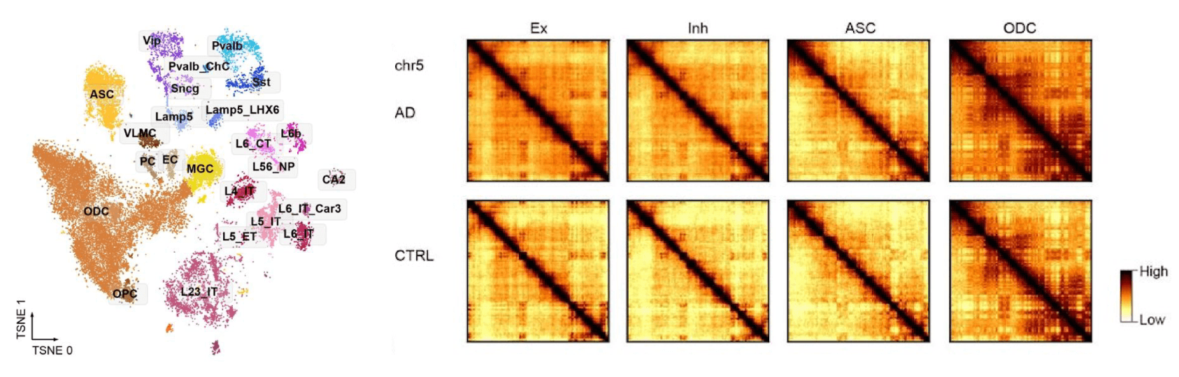
Figure 1. Cell type identification and chromosomal epigenome erosion in Alzheimer's disease brain
(Left) t-SNE projection of single-nucleus multi-omic data from postmortem entorhinal cortex, with each point representing an individual nucleus colored by its assigned cell type cluster (e.g., excitatory neuron subtypes, inhibitory neuron subtypes, astrocytes (ASC), oligodendrocytes (ODC), oligodendrocyte precursor cells (OPC), microglia (MGC), and others). Clustering was performed using methylation data obtained via single-nucleus methyl-3C sequencing (sn-m3C-seq).
(Right) Chromosomal epigenome erosion occurs in multiple cell types. Chromatin contact maps for excitatory neurons (Ex), inhibitory neurons (Inh), astrocytes (ASC), and oligodendrocytes (ODC) are shown for Alzheimer's disease (AD, top) and control (CTRL, bottom) samples across chromosome 5, as a representative example. Darker regions along the diagonal reflect stronger local chromatin contacts, while the overall loss of contrast and increased diffuse signal in AD samples relative to CTRL reflects a genome-wide erosion of chromatin domain structure. This pattern is observed consistently across cell types, indicating that chromosomal epigenome erosion is a shared feature of AD pathology rather than one confined to a single lineage.
Resource: Wang, B.-A., et al. (2023). Epigenome erosion in Alzheimer's disease brain cells and induced neurons. bioRxiv. https://doi.org/10.1101/2023.10.15.562394

(Click image to enlarge)
Figure 2. The Tle4 gene locus shows how five layers of molecular data come together to switch a gene on or off.
Using the Tle4 gene as an example, this figure shows how DNA methylation and 3D genome folding align with RNA expression and chromatin accessibility data. The four surrounding t-SNE plots color individual brain cells by each of these signals, revealing that where the gene is active (RNA and chromatin accessibility high) DNA methylation tends to be low, and vice versa, a pattern known as negative correlation, since methylation typically silences genes. The two triangular heatmaps in the center are 3D chromatin contact maps, showing that DNA folds differently near Tle4 in neurons where the gene is on ("L6 CT CTX Glut") versus off ("Pvalb GABA"), pointing to a physical loop structure that may help control the gene's activity. The genome browser tracks below translate this same story into a linear gene map, and the zoomed-in panel highlights a region upstream of the gene where multiple data types converge on the same candidate regulatory elements. Altogether, the figure illustrates how combining these different molecular signals in the same cells can reveal exactly how a gene gets switched on in one cell type and kept off in another.
Resource: Liu, H., et al. (2023). Single-cell DNA methylome and 3D multi-omic atlas of the adult mouse brain. Nature, 624(7991), 366–377. https://doi.org/10.1038/s41586-023-06805-y
Arima 3C for Single Cell Kit FAQs
No. The Arima 3C Kit provides the proximity-ligation chemistry required for single-cell 3C experiments. It is not a complete single-cell Hi-C workflow. Users must combine the kit with a published upstream crosslinking method, such as sn-m3C-seq or Dip-C, followed by a downstream single-nucleus sorting and library preparation workflow. The kit is designed for researchers developing or adapting custom single-cell 3D genomics workflows.
The kit requires crosslinked cells or nuclei containing approximately 250 ng–2.5 µg of DNA per reaction. The sample pellet for each reaction should occupy no more than 20 µL and should be free of residual liquid before starting the protocol.
The Arima 3C for Single Cell Kit User Guide includes direct crosslinking protocols for sn-m3C-seq (single-nucleus methyl-3C-seq) and Dip-C. After Arima 3C proximity ligation, users follow the appropriate published workflow for nuclei staining, sorting, and single-cell or single-nucleus library preparation.
The Arima 3C workflow includes two quality control checkpoints: Digestion QC and Ligation QC. A 10 µL aliquot is collected after restriction digestion and another after proximity ligation. These aliquots are reverse-crosslinked and purified to assess fragment-size distribution, helping users evaluate digestion and ligation efficiency before sequencing.
Yes. For bulk 3C-seq, users proceed after proximity ligation to reverse crosslinking and purification using the steps provided in the Arima 3C for Single Cell Kit User Guide. The resulting material can then be used with a preferred bulk 3C library preparation workflow.
If you need assistance at any time, please call or send an e-mail to Active Motif Technical Service at one of the locations listed below.
North America
Email: [email protected]
Phone: Toll Free - 877 222 9543
Phone: Direct - 760 431 1263
Fax: 760 431 1351
Europe
Email: [email protected]
Phone: Direct: +32 (0)2 653 0001
Fax: +32 (0)2 653 0050
Japan
Email: [email protected]
Phone: +81 (0)3 5225 3638
Fax: +81 (0)3 5261 8733
China
Email: [email protected]
Phone: (86)-21-20926090
Arima 3C Kit Publications
Reference Protocols:
- Lee D.-S. et al. Simultaneous profiling of 3D genome structure and DNA methylation in single human cells. Nature Methods 16, 999–1006 (2019). https://doi.org/10.1038/s41592-019-0547-z (sn-m3C-seq)
- Tan L. et al. Three-dimensional genome structures of single diploid human cells. Science 361, 924–928 (2018). https://doi.org/10.1126/science.aat5641 (Dip-C)
Arima 3C for Single Cell Kit Documents
You might also be interested in:
| Name | Format | Cat No. | Price | |
|---|---|---|---|---|
| Arima 3C for Single Cell Kit | 8 rxns | 53235 | $4,345 | Add to Cart |