THIS PRODUCT IS DISCONTINUED
Fast Activated Cell-based ELISA (FACE™) Kits provide a simple, sensitive method for detecting protein phosphorylation directly in the cell, without making extracts or performing electrophoresis and membrane blotting. These 96-well, high-throughput assays are available in both colorimetric and chemiluminescent formats for over 20 different targets (see list at right). For complete details, click the FACE™ Method tab below.
FACE JAK1 Kits provide 96 rxns each of 2 antibodies that enable you to monitor and compare the levels of both phosphorylated and total JAK1 (Janus Kinase 1). The phospho-JAK1 antibody recognizes JAK1 only when phosphorylated at Tyr1022 and Tyr1023. The total-JAK1 antibody recognizes JAK1 regardless of its phosphorylation state. Click the JAK1 Info tab below for data and more information.
| Name | Format | Cat No. | Price | |
|---|---|---|---|---|
| FACE™ JAK1 | 1 x 96 rxns | 48185 | Discontinued | |
| 5 x 96 rxns | 48685 | Discontinued | ||
| FACE™ JAK1 Chemi | 1 x 96 rxns | 48285 | Discontinued | |
| 5 x 96 rxns | 48785 | Discontinued | ||
| FACE™ JAK1 Manual |
| FACE™ Profile |
| Cell Biology Products Brochure |
| IsoCyte™ Application Note – Phospho-Protein Detection |
| MSDS: Sodium Azide |
| MSDS: Sulphuric Acid |
| MSDS: Thimersol |
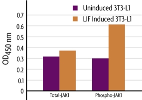
Figure 1: Measurement of phosphorylated and total JAK1.
3T3-L1 cells were cultured in 96-well plates for 48 hours and converted to adipocytes. Cells were then serum-starved for 16 hours, treated with 20 ng/ml LIF for 10 minutes and fixed. Total and phospho JAK1 were each assayed in triplicate using the phospho and total JAK1 antibodies included in the FACE JAK1 Kit. Data was plotted after correction for cell number (performed through use of Crystal Violet).
Antibody Specificities
The phospho-JAK1 antibody is specific for phosphorylated JAK1 and was raised against a synthetic phospho-peptide corresponding to residues surrounding Tyr1022 and Tyr1023 of human JAK1. This antibody recognizes JAK1 when phosphorylated at these sites and does not cross-react with related kinases. The total-JAK1 antibody recognizes JAK1 proteins regardless of the phosphorylation state.
JAK1 Overview
The Janus family of tyrosine kinases (JAK) includes JAK1, JAK2, JAK3 and Tyk2. This family associates with a variety of cytokine receptors and is activated by ligand binding. Upon ligand binding to cytokine receptors, JAKs phosphorylate themselves and their associated receptors. This provides multiple binding sites for signaling proteins containing SH2 or other phospho-tyrosine-binding domains, including STATs, Shc, insulin receptor substrates and FAK. Thus, JAK kinases provide important links between cytokine/hormone receptors and downstream effector proteins, and efficient study methods are in high demand.
The FACE™ Method
In FACE, cells are cultured in 96-well plates and stimulated to induce the pathway of interest. Following stimulation, the cells are fixed rapidly, which preserves activation-specific protein modifications. Each well is then incubated with a primary antibody specific for the activated protein of interest. Subsequent incubation with secondary HRP-conjugated antibody and developing solution provides a colorimetric or chemiluminescent readout that is quantitative and reproducible (Figure 1). The number of cells in each well can be normalized easily with the provided Crystal Violet solution. FACE Kits also contain primary antibody specific for the native inactive protein, so you can monitor both native and activated protein levels in the same experiment. FACE eliminates cellular extractions, radioactive kinase assays, time-consuming Westerns and inefficient epitope interactions that occur on membranes. FACE is a highly sensitive high-throughput assay designed for detecting activated proteins within mammalian cells.
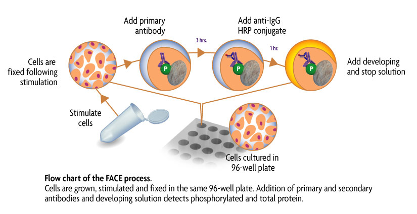
Figure 1: Flow chart of the FACE process.
Flow chart of the FACE in cell Western method that uses a cell based ELISA to measure the levels of the native and phospho forms of signaling proteins and kinases that are activated by phosphorylation.
Contents & Storage
Two (or ten) 96-well plates for culturing cells, 96 (or 5 x 96) rxns each of two primary antibodies (1 phospho-specific, 1 specific for native protein), HRP-conjugated secondary antibody, Quenching Solution, 1X Antibody Blocking Buffer, 1X Antibody Dilution Buffer, 10X PBS, 10% Triton X-100, 1% SDS Solution, Developing and Stop Solutions, and Crystal Violet Cell Quantification Solution. Storage conditions vary from room temperature to -20°C, see manual for details. All reagents are guaranteed stable for 6 months when stored properly.