Histone H3.3K27M antibody (pAb)
| RRID: AB_2793773 |
| Catalog No: 61803 | Format: 100 µl | $500 | Buy |
| Catalog No: 61804 | Format: 10 µl | $125 | Buy |
Request a quote for a bulk order Request Quote
Product Review Submit a Review
Applications

Application Notes
Applications Validated by Active Motif:
Western Blot: 1 - 2 µg/ml
Immunogen
This antibody was generated using a peptide containing a methionine substitution at lysine 27 of human Histone H3.
Buffer
Purified IgG in PBS with 30% glycerol and 0.035% sodium azide. Sodium azide is highly toxic.
View our Guide to Histone Modifications and Biological Function.
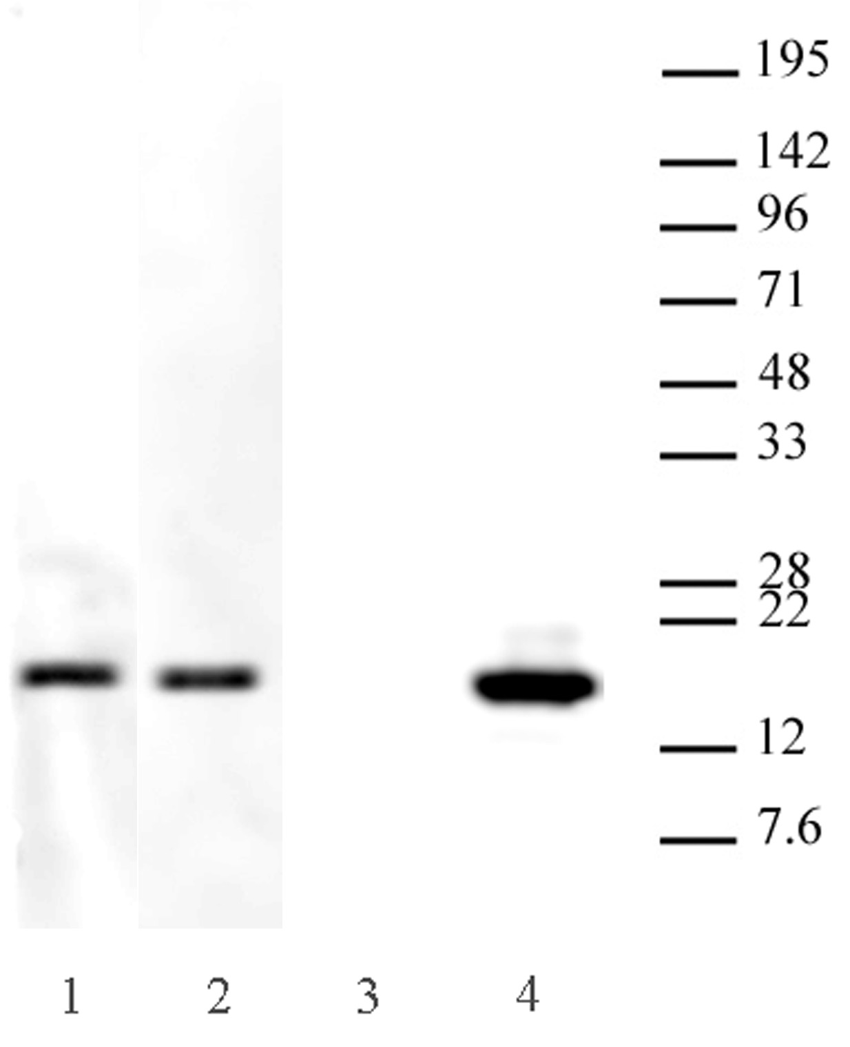
Western blot analysis of antibody specificity for Histone H3.3K27M
0.1 µg per lane of Recombinant Human Histone H3.3 Cat. No. 31295 (Lanes 1 & 3) or Recombinant Human Histone H3.3K27M Cat. No. 31552 (Lanes 2 & 4) were probed with AbFlex® Histone H3.3 antibody (rAb), Cat. No. 91191 at 2 µg/ml (Lanes 1 & 2) or Histone H3.3K27M antibody (pAb) at 0.05 µg/ml.
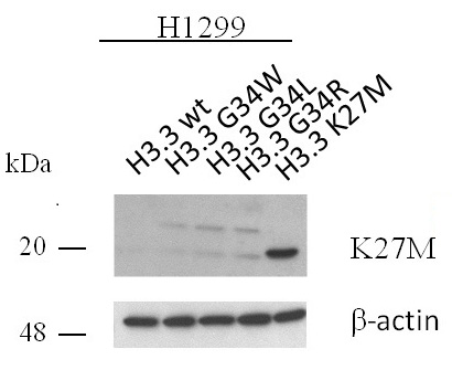
Western blot of Histone H3.3K27M pAb.
H1299 cell extracts transfected with human Histone H3.3 wild-type (wt) or various Histone H3.3 single amino acid substitutions were probed with Histone H3.3K27M antibody (pAb) at 2 µg/ml. Beta-actin was used as a loading control.
Background
Histone H3 is one of the core components of the nucleosome. The nucleosome is the smallest subunit of chromatin and consists of 146 base pairs of DNA wrapped around an octamer of core histone proteins (two each of H2A, H2B, H3 and H4). Histone H1 is a linker protein, present at the interface between the nucleosome core and DNA entry/exit points.Histone H3.1 and Histone H3.3 are the two main Histone H3 variants found in plants and animals. They are known to be important for gene regulation. Histone H3.1 and H3.3 have been shown to demonstrate unique genomic localization patterns thought to be associated with their specific functions in regulation of gene activity. Specifically, Histone H3.1 localization is found to coincide with genomic regions containing chromatin repressive marks (H3K9me3, H3K27me3 and DNA methylation), whereas Histone H3.3 primarily colocalizes with marks associated with gene activation (H3K4me3, H2BK120ub1, and RNA pol II occupancy). Deposition of the Histone H3.1 variant into the nucleosome correlates with the canonical DNA synthesis-dependent deposition pathway, whereas Histone H3.3 primarily serves as the replacement Histone H3 variant outside of S-phase, such as during gene transcription.Histone H3.3 point mutations (K27 and G34) are present in 1/3 of pediatric glioblastomas. Up to 78% of diffuse intrinsic pontine gliomas (DIPGs) carries K27M and 36% of non-brainstem gliomas carries either K27M or G34R/V oncohistone mutations. H3.3K27M expression synergizes with p53 loss and PDGFRA activation in neural progenitor cells derived from human embryonic stem cells, resulting in neoplastic transformation.
Storage
Some products may be shipped at room temperature. This will not affect their stability or performance. Avoid repeated freeze/thaw cycles by aliquoting items into single-use fractions for storage at -20°C for up to 2 years. Keep all reagents on ice when not in storage.
Guarantee
This product is guaranteed for 12 months from date of receipt.
This product is for research use only and is not for use in diagnostic procedures.
Application Key
- ChIP = Chromatin Immunoprecipitation;
- ChIP = ChIP Sequencing;
- CUT&RUN = Cleavage Under Targets and Release Using Nuclease;
- CUT&Tag = Cleavage Under Targets and Tagmentation;
- DB = Dot Blot;
- ELISA = Enzyme-linked Immunosorbent Assay;
- EMSA = Electrophoretic Mobility Shift Assay
- FC = Flow Cytometry;
- ICC = Immunocytochemistry;
- IF = Immunofluorescence;
- IHC = Immunohistochemistry;
- IP = Immunoprecipitation;
- MeDIP = Methyl-DNA Immunoprecipitation;
- NEU = Neutralizing;
- WB = Western Blotting


