Cart
 Products

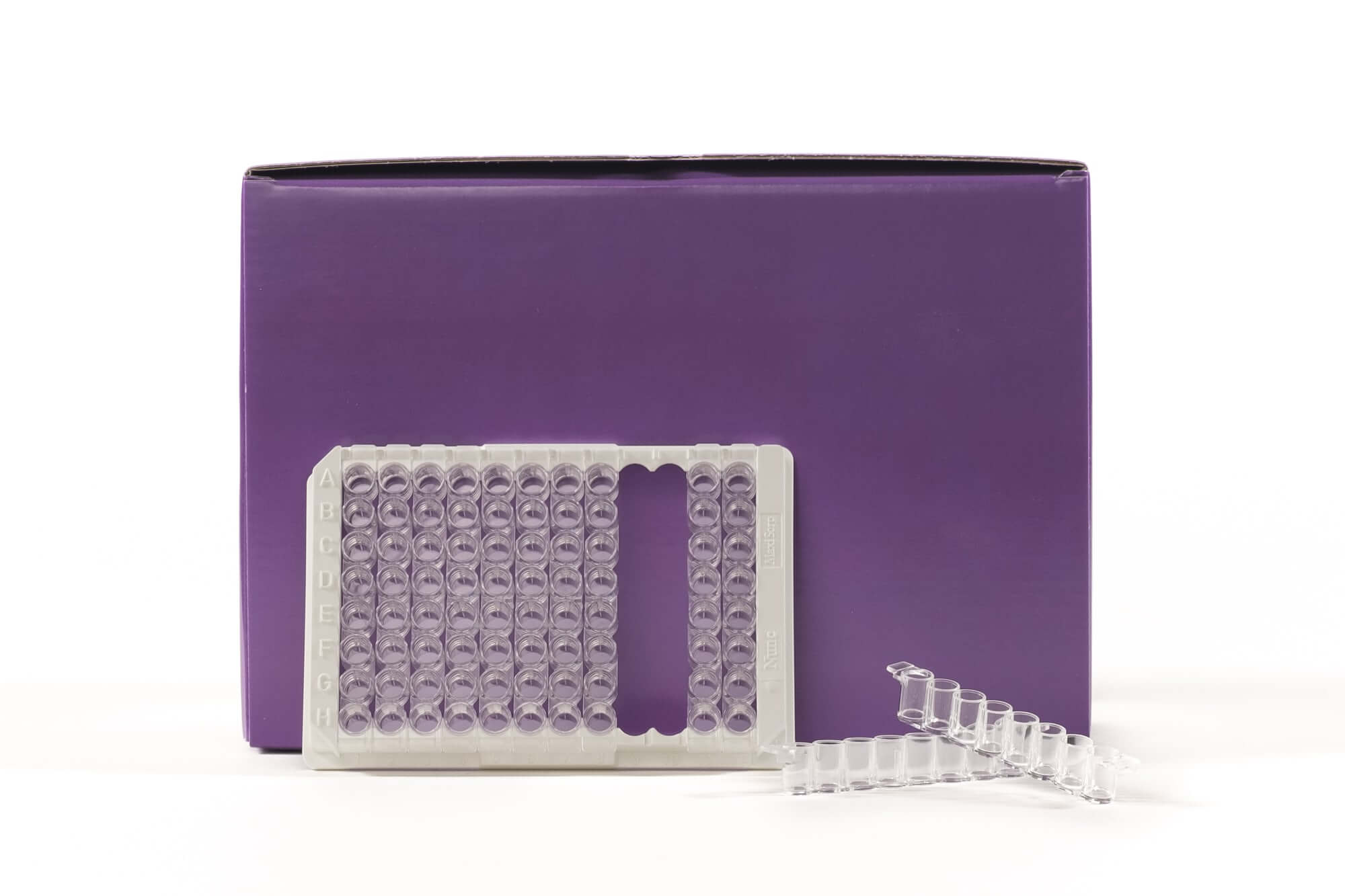
TransAM® Kits are DNA-binding ELISAs that facilitate the study of transcription factor activation in mammalian tissue and cell extracts. Assays are available for over 40 different targets (see the list at right). Each kit includes a 96-stripwell plate in which multiple copies of a specific double-stranded oligonucleotide have been immobilized. When nuclear or whole-cell extract is added, activated transcription factor of interest binds the oligonucleotide at its consensus binding site and is quantified using the included antibody, which is specific for the bound, active form of the transcription factor being studied. For complete details, click the TransAM® Method tab below.
TransAM® MyoD Transcription Factor ELISA Kits
TransAM MyoD Kits provide everything needed to study activated Myogenic Differentiation 1 (MyoD), including a positive control extract. The MyoD Kit can be used with human, mouse and rat extracts. Click the MyoD Info tab below for data and more information; kit manuals can be downloaded under the Documents tab.
THIS PRODUCT IS DISCONTINUED
| Name | Format | Cat No. | Price | |
|---|---|---|---|---|
| TransAM® MyoD | 1 x 96 rxns | 47196 | Discontinued | |
| 5 x 96 rxns | 47696 | Discontinued | ||
| TransAM® MyoD Manual |
| Gene Regulation Products & Services Brochure |
| Tools for Disease Research |
| MSDS: Sodium Azide |
| MSDS: Sulphuric Acid |
| MSDS: Thimersol |
MyoD Transcription Factor Info
Myogenic differentiation1 (MyoD), also known as myogenic factor 3 (Myf-3) is a transcription factor belonging to the myogenic factors family that contains a basic helix-loop-helix (bHLH) DNA-binding domain. The MyoD transcription factor is the central target in the signaling pathways that regulate muscle development. Although MyoD family members have been studied for decades in several muscle cell systems, the overall muscle differentiation program is still a strong area of focus to understand muscle decay.

Figure 1: Monitoring MyoD activity with the TransAM MyoD Kit:
Increasing amounts of nuclear extracts from confluent C2C12 cells (filled circles) and from NIH3T3 cells (open circles) are tested for MyoD activity using the TransAM MyoD Kit.
The TransAM® transcription factor ELISA advantage
Historically, transcription factor studies have been conducted using gelshift, Western blot and reporter plasmid transfections, which are time-consuming, do not allow for high-throughput and provide only semi-quantitative results. TransAM assays are up to 100 times more sensitive than gelshift techniques, and can be completed in less than 5 hours. Because TransAM is an ELISA-based assay*, there is no radioactivity, and the high-throughput stripwell format enables simultaneous screening of 1-96 samples. Inconsistencies due to variable reporter plasmid transfections are eliminated, along with the need to construct stable cell lines.
Why use TransAM® transcription factor ELISAs?
- Up to 100-fold more sensitive than gelshift assays
- Eliminates the use of radioactivity and the need to run gels
- Results in less than five hours
- Colorimetric readout enables easy, quantitative analysis with spectrophotometry at 450 nm
- 96-stripwell format enables both high and low throughput
How TransAM® transcription factor ELISAs work
The TransAM format is perfect for assaying transcription factor binding to a consensus-binding site. TransAM Kits contain a 96-stripwell plate to which the consensus-binding site oligo has been immobilized. Activated nuclear extract is added to each well and the transcription factor of interest binds specifically to this bound oligonucleotide. A primary antibody specific for an epitope on the bound and active form of the transcription factor is then added followed by subsequent incubation with secondary antibody and Developing Solution to provide an easily quantified, sensitive colorimetric readout (Figure 1).
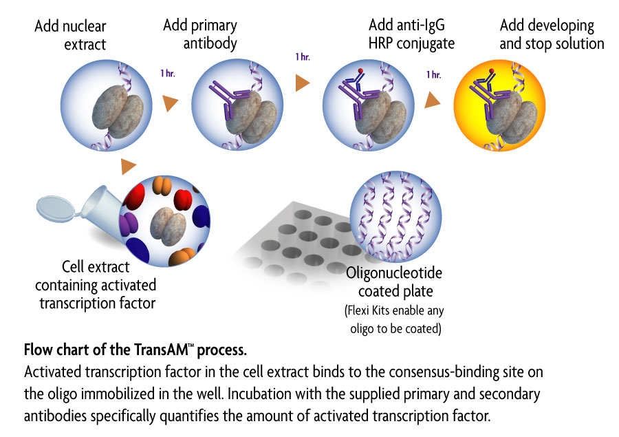
Figure 1: Flow chart of the TransAM process.
Activated transcription factor in the cell extract binds to the consensus-binding site on the oligo immobilized in the well. Incubation with the supplied primary and secondary antibodies specifically quantifies the amount of activated transcription factor.
* Technology covered under EAT-filed patents and licensed to Active Motif. Use of TransAM in NFκB-related drug discovery may be covered under U.S. Patent No. 6,150,090 and require a license from Ariad Pharmaceuticals (Cambridge, MA, USA).
Contents & Storage
One or five MyoD 96-well assay plate(s) with plate sealer(s), MyoD primary antibody, HRP-conjugated secondary antibody, wild-type and mutated oligonucleotides, C2C12 nuclear extract (positive control), Dithiothreitol, Protease Inhibitor Cocktail, poly[d(I-C)], Lysis, Binding, 10X Washing and 10X Antibody Binding Buffers, and Developing and Stop Solutions. Reagent storage conditions vary from room temperature to -80°C, see manual for details. All reagents are guaranteed stable for 6 months when stored properly.
View publications that use Active Motif TransAM® products here. Enter the product number(s) to see publications & applications that use this TransAM® product.
1. TransAM Assay Basics & Kit Selection
All TransAM kits are compatible with human samples, while some kits also cross-react with mouse or rat samples. Species cross-reactivity varies by kit.
For species compatibility information, refer to the Kit Performance and Benefits section of the TransAM kit manual. Each kit manual provides the specific cross-reactivity information for that assay.
Each TransAM kit is validated only for the species listed in the Kit Performance and Benefits section of its manual. Use with other species has not been validated by Active Motif.
Two things determine whether a kit may work in another species: the transcription factor must bind the consensus sequence coated on the plate, and the kit’s primary antibody must recognize that species’ protein. DNA-binding domains and consensus sites are often well conserved, so antibody cross-reactivity is usually the limiting factor.
Before testing a new species, compare your target protein sequence with the human sequence in the region recognized by the kit antibody.
Both colorimetric and chemiluminescent TransAM kits measure transcription factor DNA-binding activity, but they use different detection methods.
Chemiluminescent TransAM assays generally provide higher sensitivity and a greater dynamic range than colorimetric TransAM assays. The appropriate format depends on the expected abundance of the transcription factor and the sensitivity required for your experiment.
TransAM family kits are designed to analyze multiple related transcription factors within a protein family. They include two 96-well plates and antibodies against multiple transcription factors in the family. Each antibody is supplied in sufficient quantity to cover one 96-well plate, providing flexibility to analyze two or more related targets.
TransAM individual kits contain one 96-well plate and sufficient antibody for that plate, allowing analysis of a single transcription factor.
The TransAM NF-kB Flexi Kit allows researchers to study NF-kB dimers that bind to specific DNA sequences.
Unlike standard TransAM assays, the Flexi format allows you to design and bind your own custom DNA oligonucleotides to the plate. This provides flexibility for investigating NF-kB binding to specific endogenous DNA sequences.
Exact immunogen sequences are proprietary, but Technical Support can usually share the host species, clonality, and general region recognized (for example, N-terminal or C-terminal) for the antibody in your kit.
This is especially useful when working with mutant, truncated, or tagged proteins, or with compounds that bind a specific region of the transcription factor. A compound that masks the antibody epitope could reduce the signal independently of any effect on DNA binding.
The current TransAM portfolio is listed on the Active Motif website. Some kits have been discontinued over time (for example, the TransAM IRF-3 mouse kit and the TransAM Sp1 kit), so please check the website or contact Technical Support for current availability and possible alternatives.
Active Motif does not currently offer custom TransAM kits or a custom DNA-binding ELISA service. Standard TransAM plates are supplied pre-coated with a fixed consensus oligonucleotide. For NF-κB, the TransAM NF-κB Flexi Kit allows you to bind your own oligonucleotide to the plate.
2. Sample Preparation & Nuclear Extracts
TransAM assays are compatible with several nuclear extraction methods:
- Active Motif's Nuclear Extract Kit (Cat. No. 40010/40410)
- The Preparation of Nuclear Extract protocol in the TransAM manual
- Your own nuclear extraction protocol, provided the lysis buffers do not contain SDS
When using your own extraction protocol, resuspend the nuclear pellet in the Complete Lysis Buffer recommended in the TransAM kit manual.
Some TransAM kits contain Lysis Buffer AM1 while others contain Lysis Buffer AM2. Follow the compatibility instructions in the TransAM manual for the specific kit you are using.
Most TransAM kits use Lysis Buffer AM1. The TransAM NF-κB and p53 kits use Lysis Buffer AM2 because these assays do not perform as well with AM1. The buffer supplied with your kit is listed in the kit manual.
If you prepare extracts with the Nuclear Extract Kit (Cat. No. 40010/40410) or your own protocol, resuspend the nuclear pellet in the Complete Lysis Buffer specified for your TransAM kit.
If your extracts were already prepared in a different lysis buffer, you can dilute them in the correct Complete Lysis Buffer, but results may not be optimal. Preparing new extracts in the recommended buffer is preferred.
The Active Motif Nuclear Extract Kit (Cat. No. 40010/40410) includes a protocol for preparing nuclear extracts from tissue.
You may also use your own tissue nuclear extraction protocol as long as the nuclear pellet is resuspended in the Complete Lysis Buffer recommended in the TransAM kit manual. Avoid SDS because it can interfere with the TransAM assay.
Monitor cell lysis using phase-contrast microscopy.
Before lysis, intact cells appear as a dark central region representing the nucleus surrounded by a less-dense cytoplasmic halo. After successful lysis, nuclei should appear as distinct dots surrounded by asymmetric cellular debris.
Monitoring cell lysis under the microscope helps determine whether the cells have been adequately lysed while preserving the nuclei.
PBMCs can be more resistant to lysis with Hypotonic Buffer than some other cell types. Monitor lysis using phase-contrast microscopy to determine whether the cells are adequately disrupted.
If lysis is insufficient, mechanical disruption can be performed using:
- A dounce homogenizer with a small-clearance pestle (0.025–0.076 mm)
- A 28–30 gauge needle to mechanically shear the sample
Active Motif offers 1 mL and 15 mL dounce homogenizers with both small- and large-clearance pestles.
Primary cells can be challenging to process because they are often fragile and may lyse easily during scraping. Sample availability may also be limited.
Monitor cell lysis under a microscope during nuclear extraction to help maximize nuclear extract yield while minimizing unnecessary sample disruption.
For cultured cells, use a dounce homogenizer with a small pestle clearance of 0.025–0.076 mm to lyse cells and release nuclei.
For tissue, use a large-clearance pestle (0.089–0.14 mm) to first homogenize the tissue into a single-cell slurry, followed by a small-clearance pestle to lyse the cells and release the nuclei.
Active Motif's 1 mL and 15 mL dounce homogenizers include both small- and large-clearance pestles.
The Active Motif Nuclear Extract Kit (Cat. No. 40010/40410) can be used with both fresh and frozen cell and tissue samples. Fresh cells or tissue are always preferred for nuclear extract preparation. Freezing and thawing can rupture some nuclei, which typically lowers nuclear extract yield and can leave some nuclear proteins in the cytoplasmic fraction. However, frozen samples can be used if necessary.
Frozen tissue
Frozen tissue can be used with the Active Motif Nuclear Extract Kit (Cat. No. 40010/40410). It will produce quality nuclear extract, but at a lower yield than fresh tissue.
Frozen cells
Cryopreserved cells could potentially be used, although this has not been validated by Active Motif.
- Freeze cells in a cryopreservation medium (for example, 90% FBS/10% DMSO) using a controlled-rate freezing container.
- After thawing, wash the cells to remove the freezing medium and confirm viability before extraction.
- If you plan to stimulate or treat the cells, do so after thawing and recovery, then prepare nuclear extracts immediately.
Freezing cells after treatment may not preserve the activation state of the transcription factor. This is especially true for factors with transient activation, such as NFAT or HIF-1. For these targets, prepare nuclear extracts from fresh cells and freeze the extracts in single-use aliquots at −80°C instead.
Activation events such as dephosphorylation, stabilization, and nuclear translocation can be lost during sample handling. Whenever possible, prepare nuclear extracts directly from freshly treated or freshly isolated cells, and freeze the extracts rather than the cells.
- Keep cells and buffers cold throughout the extraction and work quickly.
- Use protease inhibitors, and phosphatase inhibitors where phosphorylation state matters. For targets with rapid proteasomal turnover, such as HIF-1α, some researchers also add a proteasome inhibitor such as MG132 (not validated by Active Motif).
- Aliquot extracts into single-use volumes and freeze immediately at −80°C.
- Confirm that your stimulus (for example, hypoxia or CoCl₂ treatment for HIF-1) induces nuclear translocation in your cell type.
If the kit’s positive control extract gives the expected signal but your samples do not, sample preparation or transcription factor activation is the most likely cause.
Avoid repeated freeze-thaw cycles by aliquoting nuclear extracts before storage.
If freeze-thawing is unavoidable, no more than two freeze-thaw cycles are recommended. Multiple freeze-thaw cycles can degrade proteins and reduce protein activity.
Thaw extracts on ice and mix by gentle pipetting. Avoid vigorous vortexing of protein samples.
If the nuclear pellet is difficult to resuspend in Complete Lysis Buffer, try the following:
- Pipet up and down several times using a large-bore pipette tip.
- Use a dounce homogenizer.
- Vortex at high speed for approximately 10 seconds.
- Extend the rocking incubation from 30 minutes to 1 hour.
- If necessary, add 5–10 µL of Complete Lysis Buffer, taking care not to excessively dilute the sample.
Some cell types naturally produce more viscous nuclear pellets and can still provide good protein yields even when the pellet does not completely dissolve.
Do not add detergent, because it may interfere with the TransAM assay.
It depends on the transcription factor being studied.
Whole-cell extracts dilute the transcription factor into a larger volume compared with nuclear extracts, so approximately two times or more protein may be required.
For transcription factors whose activity is regulated by nuclear-cytoplasmic localization, whole-cell extracts may also affect the functional interpretation of the assay. Nuclear extracts are generally preferred when the assay is intended to measure nuclear transcription factor DNA-binding activity.
Cytoplasmic extracts are not recommended for TransAM assays.
TransAM assays measure protein-DNA binding, which occurs in the nucleus. Signal from a cytoplasmic extract may therefore not represent physiologically relevant transcription factor activity.
Cytoplasmic extracts can also be more dilute than nuclear extracts and may contain detergents or other components that interfere with the assay.
3. Sample Input, Protein Quantification & Assay Setup
The minimum detectable amount varies between TransAM kits and is specified in the individual kit manual.
For example, some TransAM NF-kB kits can detect DNA binding from as little as 0.5 µg of nuclear extract. Using an approximate yield of 20 µg nuclear extract per 1 million cells, 0.5 µg of nuclear extract corresponds to approximately 25,000 cells.
Always check the detection limit specified for the specific TransAM kit being used.
The optimal protein input should be determined empirically because the appropriate amount depends on the sample and the specific TransAM assay.
A small protein titration is recommended. For nuclear extracts, a starting range of approximately 5, 10, and 20 µg per well (confirm with specific kit manual) can be used to identify an appropriate input level. Whole-cell extracts may require more protein.
The goal is to ensure that both treated and control samples produce signals within the linear range of the assay.
Active Motif recommends using a Bradford assay for nuclear extract protein quantification.
Standard BCA assays are not compatible with reducing agents such as DTT, which is present in the Complete Lysis Buffer. Reducing agents interfere with the BCA chemistry and can make protein concentrations appear higher than they are. Bradford assays are generally more tolerant of reducing agents but can be affected by detergents, so samples should be diluted before measurement.
When using the Active Motif Nuclear Extract Kit (Cat. No. 40010/40410), a 1:50 to 1:250 dilution in water is recommended. Prepare the protein standards in Complete Lysis Buffer diluted the same way as your samples, or at minimum include a blank of diluted Complete Lysis Buffer. The Nuclear Extract Kit manual also includes a protocol for generating a BSA standard curve.
Other protein quantification assays that are compatible with reducing agents and detergents are commercially available and could potentially be used. However, Active Motif has not validated these assays with TransAM kits or the Nuclear Extract Kit.
For nuclear extract preparation, Active Motif suggests starting with approximately:
- 8.8 × 10⁶ cells, or
- 45 mg tissue
For general estimation, a tissue conversion factor of approximately 2 × 10⁵ cells per mg tissue can be used.
Using the Active Motif Nuclear Extract Kit (Cat. No. 40010/40410), approximately 8.8 × 10⁶ cells (about the number of HeLa cells in a confluent 100 mm dish) typically yield:
- Nuclear extract: approximately 150–250 µg of protein at 3–5 mg/mL
- Cytoplasmic extract: approximately 0.5–1 mg at 1–2 mg/mL
- Whole-cell extract: approximately 1.2–2.4 mg at 4–8 mg/mL
Actual yields vary with cell or tissue type and extraction efficiency.
SDS and sodium azide in the cell lysis buffer can interfere with TransAM assays. These components can inhibit binding interactions and may make the sample incompatible with the assay.
When preparing nuclear extracts, use the lysis buffer recommended in the TransAM kit manual and avoid SDS-containing buffers.
4. Running & Interpreting the TransAM Assay
The recommended development time is provided in the technical data sheet (TDS) for each lot and should be used as a starting guideline.
Because color development can vary between samples, monitor the color development of your specific samples rather than relying solely on the recommended incubation time. The TransAM color chart can be used as an additional reference when evaluating colorimetric assay development.
Be sure to bring the Developing Solution to room temperature before use and keep the plate protected from light during development.
OD450 is the primary absorbance measurement, while OD655 serves as a reference wavelength.
Subtracting the reference absorbance from the primary measurement helps correct for variation caused by factors such as differences between wells and detection equipment.
The corrected signal is calculated as:
OD450 − OD655
Many spectrophotometers and plate-reader software programs can perform this correction automatically.
At minimum, TransAM assays should be performed in duplicate to evaluate technical variability.
If the assay shows high variability between replicates, running triplicates is recommended.
When presenting the results, plotting the data with standard error can help show the variability between technical replicates.
A standard curve is not required for most TransAM experiments. TransAM assays are typically used to compare relative transcription factor DNA-binding activity between samples, such as treated and untreated, and a relative comparison is often more informative than an absolute concentration.
A standard curve can be useful if you want to estimate the amount of active transcription factor in your samples.
The wild-type consensus oligonucleotide competes with the plate-bound probe for transcription factor binding and should reduce the signal, while the mutated oligonucleotide should have little or no effect. Together they demonstrate that the signal is specific.
The degree of inhibition varies between kits. For some kits, such as TransAM c-Myc, the expected inhibition with the wild-type oligonucleotide is moderate (approximately 20–40%). Check the manual and lot-specific TDS for your kit.
This can vary depending on the lot and spectrometer used. Always run the positive control extract supplied with the kit.
This has not been validated by Active Motif. Some researchers have detected tagged recombinant proteins (for example, FLAG-tagged) bound to the plate using their own anti-tag primary antibody and a matching HRP-conjugated secondary antibody. Antibody dilutions need to be optimized, and appropriate controls should be included, such as wells without protein and the wild-type and mutated competitor oligonucleotides.
5. Troubleshooting & Assay Optimization
Low TransAM assay signal can result from several factors, including insufficient protein input or low levels of active transcription factor in the sample.
Start by checking:
- Protein input
- Nuclear extraction efficiency
- Sample preparation
- Transcription factor abundance or activation state
- Whether the sample signal falls within the assay's detectable range
A small protein titration can help determine whether increasing sample input improves the signal while maintaining the assay within its linear range. Always include the positive control extract in at least 1 well per run.
Common sources of well-to-well variability include:
- Developing Solution not equilibrated to room temperature before use.
- Pipetting error. Prepare a master mix of each sample in Complete Lysis Buffer and pipette all replicates from the same tube.
- Adding undiluted sample directly to the well. Mix the sample with Complete Lysis Buffer before adding it, because binding incubations are performed without agitation.
- Residual wash buffer. Tap the inverted plate on paper towels to remove all liquid.
- Sloshing the plate, which can cross-contaminate wells. Swirl gently by hand once before incubations.
- 10X buffers not diluted to 1X, or buffers not mixed thoroughly.
- Developing Solution contaminated with Stop Solution. Use separate, clean reservoirs for each reagent.
- Bubbles in the wells at the time of reading, or light exposure during development.
- Plate exposed to moisture, or extracts stored at −20°C or freeze-thawed more than twice.
- Plate reader wells that do not read correctly. Test the reader or use a different instrument.
If variability persists, run triplicates and contact Technical Support with your raw data, kit lot number, and plate reader settings.
For troubleshooting information specific to your TransAM kit, refer to the product manual for that assay. The manual contains kit-specific instructions and recommendations for sample preparation, assay performance, and troubleshooting.
6. Kit Storage, Components & Ordering
TransAM plates are very sensitive to moisture. Return any unused wells to the original foil pouch with the desiccant, seal the pouch tightly (taping it closed works well), and store as indicated in the manual. Check that the pouch is not torn. Plates exposed to moisture, or stored outside the pouch, can give anomalous results.
Storage temperatures for each component are listed in the kit manual. Components have different requirements, and some antibodies should not be frozen, so do not store the entire kit at a single temperature unless the manual specifies it.
Nuclear extracts should be stored in single-use aliquots at −80°C. Storage at −20°C is not recommended.
Contact Technical Support as soon as possible with your order number, kit lot number, and a description or photo of the shipment condition. Store the components at the recommended temperatures in the meantime, and check with us before using the kit.
Contact Technical Support with your catalog number and kit lot number. SDS documents for TransAM kit components are also available on request.
Some individual components can be quoted on request. Contact Technical Support with your catalog number and the components and quantities you need. When multiple units of the major components (plate, antibodies, and buffers) are needed, purchasing the complete kit is usually more cost-effective.