Active Motif's fluorescent secondary antibody conjugates provide you with an array of choices for immunofluorescent detection of primary antibodies. Our secondary antibodies are conjugated to a number of high-quality dyes, including the Chromeo™ line of fluorescent dyes. We also offer ATTO dye conjugates that have been maximally cross-adsorbed against IgG's of a variety of species to eliminate background caused by non-specific binding. Chromeo 488, Chromeo 505, Chromeo 494 and the ATTO STED dyes have been certified by Leica Microsystems for STimulated Emission Depletion (STED) microscopy, allowing a spatial resolution of 50 - 70 nm.
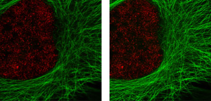
Figure 1: Active Motif's primary antibodies and fluorescent secondary antibodies in confocal and STED microscopy.
HeLa cells were stained with alpha Tubulin mouse mAb (Clone 5-B-1-2) and ATTO 647N (STED/GSD) Goat anti-mouse IgG (Catalog No. 15038). Histone H3 was stained with Histone H3 trimethyl Lys4 rabbit polyclonal antibody (Catalog No. 39159) and the Chromeo 488 Goat anti-rabbit IgG (Catalog No. 15041) secondary antibody. The confocal (left) and STED (right) images are courtesy of Leica Microsystems, Germany.
Chromeo 488, Chromeo 505, Chromeo 546, ATTO 532 (GSD), ATTO 647N (STED/GSD) and the Rhodamine 6G (GSD) secondary antibody conjugates are recommended by Leica Microsystems for high-resolution GSDIM microscopy, using the novel SR GSD microscope. With this novel high resolution technique a resolution of about 20 nm can be achieved.
Click on antibody conjugate below to see its complete information. To see all of all our antibodies, please go to the All Antibodies page.
Chromeo™ Dyes are bright fluorescent labels that replace Alexa Fluor*, DyLight* or Cy*-dyes. They are compatible with most excitation sources including diode lasers, LEDs, tungsten lamps and xenon arc lamps. The dyes have been conjugated to high-quality secondary antibodies by an optimized conjugation method, including subsequent purification from interfering substances.
Chromeo™ Dye Secondary Antibody Conjugate advantages
- High intensity
- Specificity under various fixation conditions
- Photostability – the combination with MAX Stain™ reagents provides optimal fluorescent stability for multiple exposures and increased exposure time
- Low background
Fluorescent Chromeo Dyes Secondary Antibody Conjugates enable sensitive and specific detection in fluorescence microscopy, high content screening, ELISA, FRET applications or flow cytometry. To ensure that you get the best results possible, we highly recommend that you use our secondary antibody conjugates together with our MAX Stain™ Immunofluorescence Tools, as those components have been formulated to optimize the performance of the Chromeo™ Dyes.
To receive more detailed information and application data about individual Chromeo dyes, simply click on the name of the Chromeo dye of your choice in the table below. To see a larger spectra image, click on the spectrum of your choice.
| Dye | Absorption | Emission | Spectra | ε L/(mol-cm) | Stokes shift |
|---|---|---|---|---|---|
| Chromeo™ 488 | 498 nm | 524 nm |  |
73,000 | 26 nm |
| Chromeo™ 494 | 489 nm | 624 nm |  |
55,000 | 135 nm |
| Chromeo™ 505 | 514 nm | 530 nm |  |
70,000 | 16 nm |
| Chromeo™ 546 | 550 nm | 567 nm |  |
98,800 | 17 nm |
| Chromeo™ 642 | 647 nm | 666 nm |  |
180,000 | 19 nm |
| ATTO 647N (STED) | 644 nm | 669 nm |  Fluorescent Dye Spectra in a new window") |
150,000 | 25 nm |
| ATTO 655 (STED) | 663 nm | 684 nm |  Fluorescent Dye Spectra in a new window") |
125,000 | 21 nm |
| ATTO 532 (GSD) | 534 nm | 560 nm |  Fluorescent Dye Spectra in a new window") |
115,000 | 26 nm |
| Rhodamine 6G (GSD) | 508 nm | 558 nm |  Fluorescent Dye Spectra in a new window") |
116,000 | 50 nm |
| Table 1: Properties of Active Motif's Fluorescent Antibody Conjugates. | |||||
Next to the spectral properties of the dye and the quality of the secondary antibody, the quality of a fluorescent conjugate is influenced by the dye-to protein ratio, the conjugation method and its purity. All Active Motif Secondary Antibody Conjugates have been prepared by an optimized conjugation protocol making the fluorescent secondaries brighter and lowering the fluorescent background. Active Motif antibody conjugates have been tested in various applications including flow cytometry and fluorescent microscopy where they have shown to work with high efficiency and specificity under multiple fixation conditions.
Active Motif Fluorescent Secondary advantages
- Unrivaled fluorescent intensity
- Low background
- Limited photobleaching
- High Specificity
- Flexibility
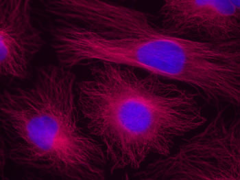
Figure 1: Chromeo 642 staining in HeLa cells.
HeLa cells were stained with alpha Tubulin mouse mAb (Clone 5-B-1-2) and Chromeo 642 Goat anti-mouse IgG (Catalog No. 15034). The nuclei have been counterstained with DAPI.
Figure 2: Chromeo 488 antibody conjugates in STED microscopy.
Nuclear pore protein-1 (NUP-1) was stained with a primary monoclonal mouse antibody and with Chromeo 488 Goat anti-mouse IgG (Catalog No. 15031) secondary antibody (left). Vimentin was stained with a primary polyclonal rabbit antibody and with Chromeo 488 Goat anti-rabbit IgG (Catalog No. 15041) secondary antibody (right). These STED images are courtesy of Leica Microsystems, Germany.
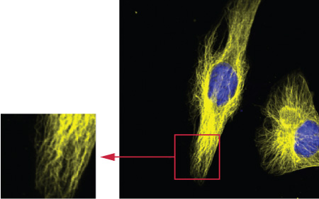
Figure 3: Tubulin staining in HeLa cells, analyzed by confocal microscopy using the 544 nm line of the HeNe laser.
HeLa cells were stained with alpha Tubulin mouse mAb (Clone 5-B-1-2) and Chromeo 546 Goat anti-mouse IgG (Catalog No. 15033).
The Fluorescent Secondary Antibodies have been cited in the following publications:
- “Molecular basis of xeroderma pigmentosum group C DNA recognition by engineered meganucleases” by Redondo et al (2008) Nature 456:107-111. (Chromeo 488 antibody conjugate)
- “The WSB1 gene is Involved in Pancreatic Cancer Progression” by Archange et al (2008) Plos ONE 3(1):e2475. (Chromeo 488 antibody conjugate)
- “The Good into the Pot, the bad into the Crop-A New Technology to Free Stem Cells from Feeder Cells” by Schneider et al (2008) Plos ONE 3(11):e3788. (Chromeo 488 antibody conjugate)
- “Dual-Color STED Microscopy at 30-nm Focal-Plane Resolution” by Meyer et al (2008) Small 4(8):1095-1100. (Chromeo 488 antibody conjugate)
- “The ABC transporter-encoding gene AFR1 affects the resistance of Cryptococcus neoformans to microglia-mediated antifungal activity by delaying phagosomal maturation” by Orsi et al (2009) FEMS yeast res. 9:301-310. (Chromeo 642 antibody conjugate)
- “p-ERK1/2 is a predictive factor of response to erythropoiesis-stimulating agents in low/int-1 myelodysplastic syndromes” by Frisan et al (2010) Haematologica 95(11):1964-1968. (Chromeo 642 antibody conjugate)
- “An Inexpensive Simple-to-Use Inverted Fluorescence Microscope: A new Tool for Cellular Analysis” by Kahle et al (2010) J.of Lab. Autom. 15(5):355-361. (Chromeo 488 antibody conjugate)
- “Direct Synthesis of Lamin A, bypassing Prelamin A processing, Causes Misshapen Nuclei in Fibroblasts but No Detectable Pathology in Mice” by Coffinier et al (2010) J Biol. Chem. 285(27):20818-20826. (ATTO 647N antibody conjugate)
- “PTPIP51 is phosphorylated by Lyn and c-Src kinases lacking dephosphorylation by PTP1B in acute myeloid leukemia” by Brobeil et al (2011) Leukemia Research 35(10):1367-1375. (Chromeo 488 antibody conjugate)
- “Role of the (Mn)superoxide dismutase of Enterococcus faecalis in the in vitro interaction with microglia” by Peppoloni et al (2011) Microbiology 157:1816-1822. (Chromeo 642 antibody conjugate)
- “In Vitro Investigation of DNA Damage Induced by the DNA Cross-Linking Agents Oxaliplatin and Satraplatin in Lymphocytes of Colorectal Cancer Patients” by Alotaibiet al (2012) J.of Cancer Therapy 3:78-89. (Chromeo 488 antibody conjugate)
- “Local palmitoylation cycles define activity-regulated postsynaptic subdomains” by Fukata et al (2013) JCB 202(1):145-161. (Chromeo 505 antibody conjugate)
- “Fast neurotransmitter release regulated by the endocytic scaffold intersectin” by Sakaba et al (2013) PNAS 110(20):8266-8273. (Chromeo 494 antibody conjugate)
Contents & Storage
The Chromeo™ conjugates are supplied as 1 mg of antibody at a concentration of 2 mg/ml. The ATTO and the Rhodamine 6G conjugates are supplied in 200 or 35 µl aliquots in PBS. For short-term storage, the conjugated antibody should be stored at 4°C protected from light. For longer-term storage, aliquot the antibody and store at -20°C. Avoid subjecting the antibody to repeated freeze-thaw cycles. These products are guaranteed for 6 months from the date of arrival.
To ensure photostability of Chromeo 488 under all experimental conditions, one aliquot of MAXfluor™ Mounting Medium for use in fluorescence microscopy is supplied with Chromeo 488 conjugates. This will provide optimal fluorescence stability and anti-fading during long-term storage of your mounted slides. MAXfluor Mounting medium should be stored at 4°C.
*All trademarks are the property of their respective owners.